File:Hilfer1990 Fig16.jpg
From Embryology
Size of this preview: 540 × 600 pixels. Other resolution: 1,200 × 1,333 pixels.
Original file (1,200 × 1,333 pixels, file size: 247 KB, MIME type: image/jpeg)
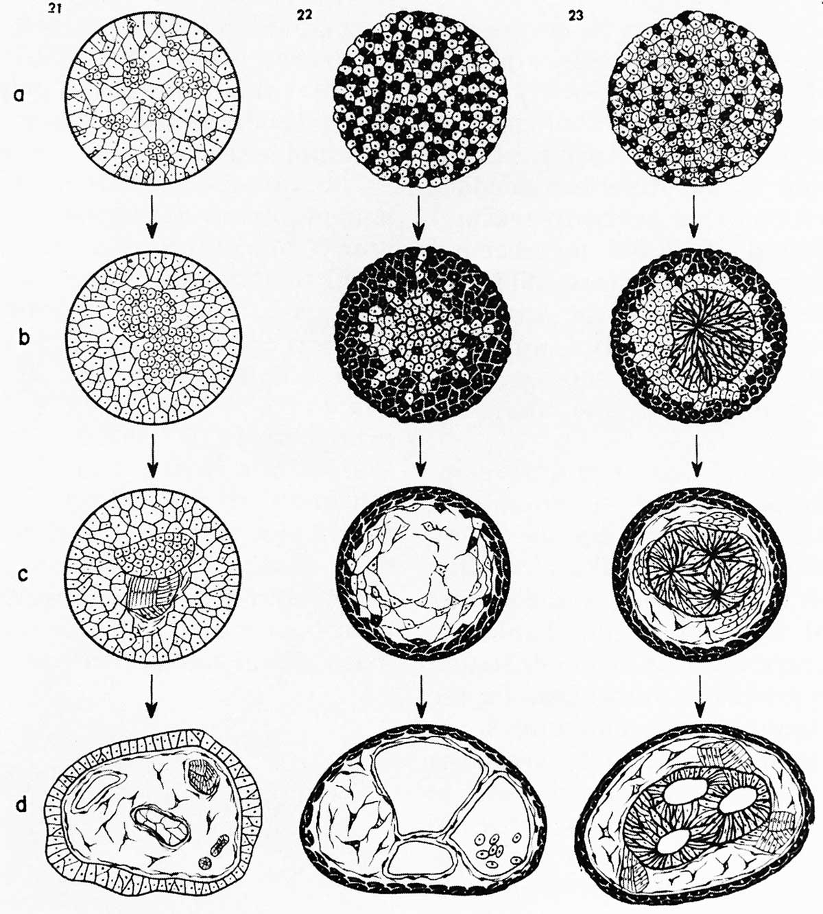
Figure 16. Townes and Holtfreter (1955) different embryonic layers in amphibians
Illustration from Townes and Holtfreter (1955) demonstrating sorting of dissociated cells from different embryonic layers in amphibians. Cells from different germ layers sorted into clumps and tended to take a position resembling that of normal embryonic development.
- Figures: Fig 1. by N. Hartsoeker 1694 | Fig 2. by M. Malpighi 1673 | Fig 3. by C.E. von Baer 1827 | Fig 4. by W. Roux 1888 | Fig 5. by H. Driesch 1892 | Fig 6. Louis Agassiz | Fig 7. Leonard W. Williams c1900 | Fig 8. by Conklin 1905 | Fig 9. by Wilson 1892 | Fig 10. by Loeb 1893 | Fig 11. by E. B. Wilson 1904 | Fig 12. by O.E. Schotte | Fig 13. by Spemann and H. Mangold 1924 | Fig 14. by S. Horstadius 1928 | Fig 15. by R. G. Harrison 1921 | Fig 16. by Townes and Holtfreter 1955
Cite this page: Hill, M.A. (2024, April 25) Embryology Hilfer1990 Fig16.jpg. Retrieved from https://embryology.med.unsw.edu.au/embryology/index.php/File:Hilfer1990_Fig16.jpg
- © Dr Mark Hill 2024, UNSW Embryology ISBN: 978 0 7334 2609 4 - UNSW CRICOS Provider Code No. 00098G
File history
Click on a date/time to view the file as it appeared at that time.
| Date/Time | Thumbnail | Dimensions | User | Comment | |
|---|---|---|---|---|---|
| current | 10:42, 28 August 2014 | | 1,200 × 1,333 (247 KB) | Z8600021 (talk | contribs) | Figure 16. Illustration from Townes and Holtfreter (1955) demonstrating sorting of dissociated cells from different embryonic layers in amphibians. Cells from different germ layers sorted into clumps and tended to take a position resembling that of nor... |
You cannot overwrite this file.
File usage
The following page uses this file: