File:Hilfer1990 Fig02.jpg
From Embryology

Size of this preview: 410 × 599 pixels. Other resolution: 1,027 × 1,500 pixels.
Original file (1,027 × 1,500 pixels, file size: 156 KB, MIME type: image/jpeg)
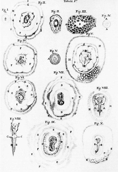
Figure 2. Illustrations from M. Malpighi (1673)
Figure 2. Illustrations from M. Malpighi (1673) of developing chicken embryos from the time of laying to 36 hours of incubation. This plate is representative of the keen observations done on early embryos during the 17th century.
- Figures: Fig 1. by N. Hartsoeker 1694 | Fig 2. by M. Malpighi 1673 | Fig 3. by C.E. von Baer 1827 | Fig 4. by W. Roux 1888 | Fig 5. by H. Driesch 1892 | Fig 6. Louis Agassiz | Fig 7. Leonard W. Williams c1900 | Fig 8. by Conklin 1905 | Fig 9. by Wilson 1892 | Fig 10. by Loeb 1893 | Fig 11. by E. B. Wilson 1904 | Fig 12. by O.E. Schotte | Fig 13. by Spemann and H. Mangold 1924 | Fig 14. by S. Horstadius 1928 | Fig 15. by R. G. Harrison 1921 | Fig 16. by Townes and Holtfreter 1955
Cite this page: Hill, M.A. (2024, April 16) Embryology Hilfer1990 Fig02.jpg. Retrieved from https://embryology.med.unsw.edu.au/embryology/index.php/File:Hilfer1990_Fig02.jpg
- © Dr Mark Hill 2024, UNSW Embryology ISBN: 978 0 7334 2609 4 - UNSW CRICOS Provider Code No. 00098G
File history
Click on a date/time to view the file as it appeared at that time.
| Date/Time | Thumbnail | Dimensions | User | Comment | |
|---|---|---|---|---|---|
| current | 10:53, 28 August 2014 | | 1,027 × 1,500 (156 KB) | Z8600021 (talk | contribs) | |
| 20:29, 23 August 2014 | 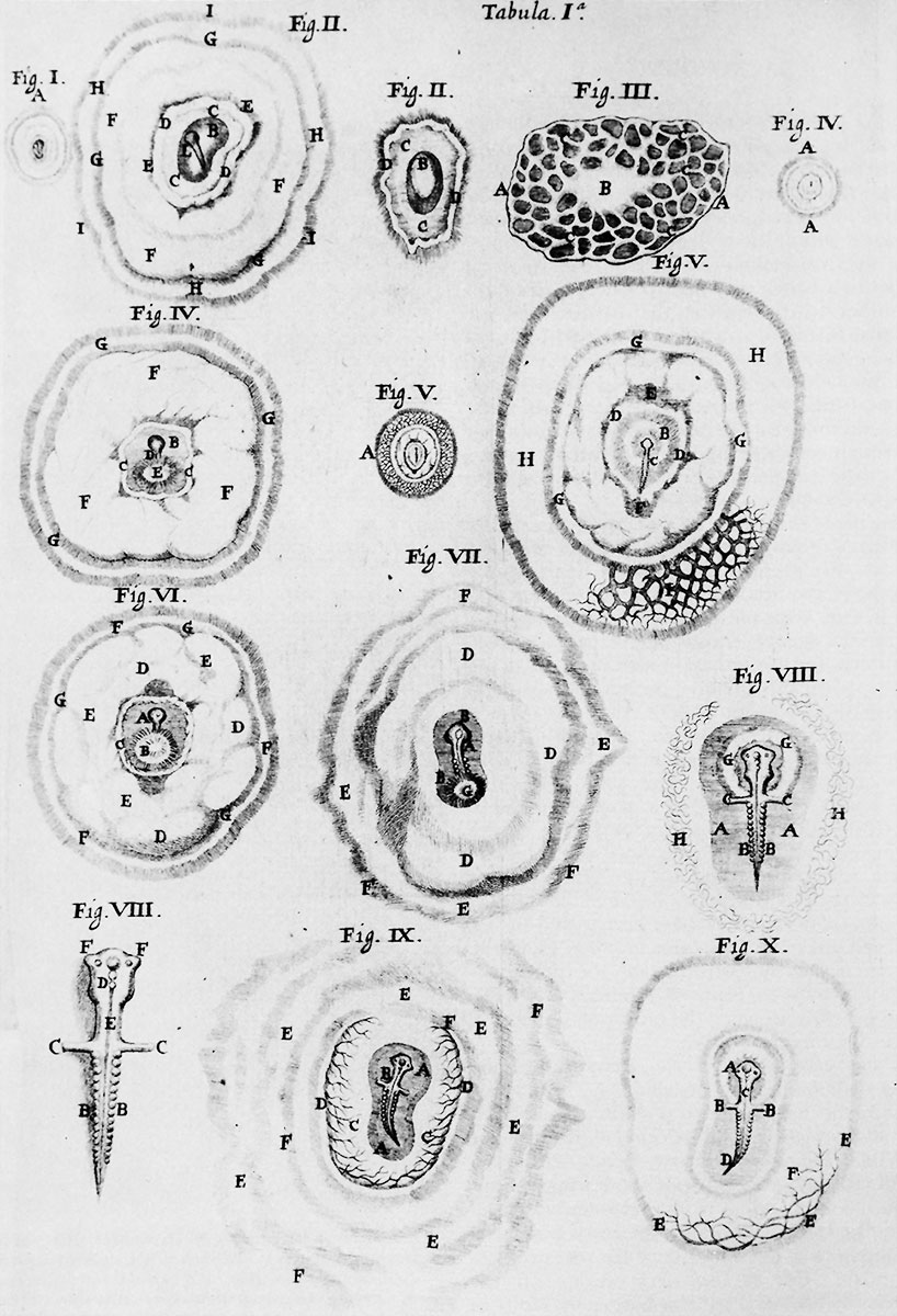 | 818 × 1,200 (204 KB) | Z8600021 (talk | contribs) | ||
| 20:27, 23 August 2014 | 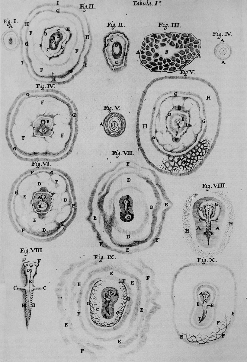 | 818 × 1,200 (215 KB) | Z8600021 (talk | contribs) | ||
| 20:26, 23 August 2014 | 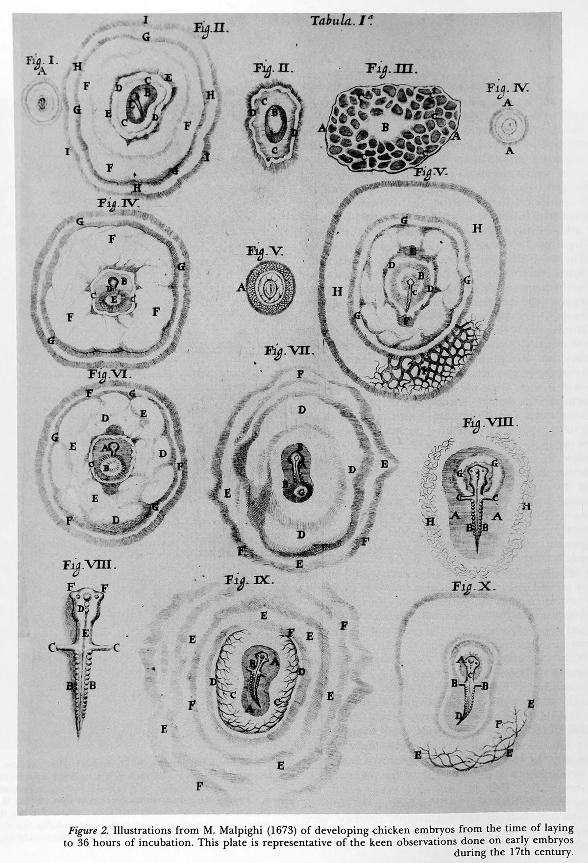 | 1,200 × 1,761 (465 KB) | Z8600021 (talk | contribs) | {{Hilfer1990figures}} |
You cannot overwrite this file.
File usage
The following page uses this file: