File:Hertwig286.jpg: Difference between revisions
From Embryology
(==Fig. 286. Frontal reconstruction of the oro-pharyngeal cavity of a human embryo (Rg of His) 11.5 mm long== neck measurement. From His, " Menschliche Bmbryonen." Magnified 12 diameters. The upper jaw is seen in perspective, the lower jaw in section. T) |
mNo edit summary |
||
| Line 1: | Line 1: | ||
==Fig. 286. Frontal reconstruction of the oro-pharyngeal cavity of a human embryo (Rg of His) 11.5 mm long== | ==Fig. 286. Frontal reconstruction of the oro-pharyngeal cavity of a human embryo (Rg of His) 11.5 mm long== | ||
neck measurement. From His, " Menschliche | neck measurement. | ||
From {{His}}, " Menschliche Embryonen." Magnified 12 diameters. | |||
The upper jaw is seen in perspective, the lower jaw in section. The posterior visceral arches are not visible from the outside, since they have moved into the depths of the cervical sinus. | The upper jaw is seen in perspective, the lower jaw in section. The posterior visceral arches are not visible from the outside, since they have moved into the depths of the cervical sinus. | ||
| Line 8: | Line 10: | ||
{{Hertwig images}} | {{Hertwig images}} | ||
[[Category:Hearing]] [[Category: | [[Category:Hearing]] [[Category:Head]] | ||
Latest revision as of 15:12, 24 June 2013
Fig. 286. Frontal reconstruction of the oro-pharyngeal cavity of a human embryo (Rg of His) 11.5 mm long
neck measurement.
From Wilhelm His (1831-1904), " Menschliche Embryonen." Magnified 12 diameters.
The upper jaw is seen in perspective, the lower jaw in section. The posterior visceral arches are not visible from the outside, since they have moved into the depths of the cervical sinus.
| Historic Disclaimer - information about historic embryology pages |
|---|
 |
Reference
Hertwig O. Text-book of the embryology of man and mammals. (1892) Translated 1901 by Mark EL. from 3rd German Edition. S. Sonnenschein, London.
Cite this page: Hill, M.A. (2024, April 19) Embryology Hertwig286.jpg. Retrieved from https://embryology.med.unsw.edu.au/embryology/index.php/File:Hertwig286.jpg
- © Dr Mark Hill 2024, UNSW Embryology ISBN: 978 0 7334 2609 4 - UNSW CRICOS Provider Code No. 00098G
File history
Click on a date/time to view the file as it appeared at that time.
| Date/Time | Thumbnail | Dimensions | User | Comment | |
|---|---|---|---|---|---|
| current | 11:56, 20 May 2011 | 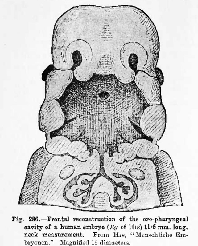 | 642 × 800 (126 KB) | S8600021 (talk | contribs) | ==Fig. 286. Frontal reconstruction of the oro-pharyngeal cavity of a human embryo (Rg of His) 11.5 mm long== neck measurement. From His, " Menschliche Bmbryonen." Magnified 12 diameters. The upper jaw is seen in perspective, the lower jaw in section. T |
You cannot overwrite this file.
File usage
The following page uses this file: