File:HertigAdams1967 fig04.jpg

Original file (1,628 × 1,714 pixels, file size: 383 KB, MIME type: image/jpeg)
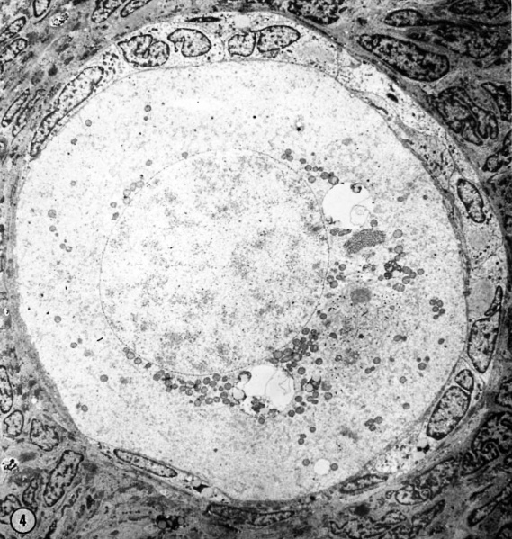
Fig. 4 A low—power survey micrograph through the mid-polar axis of an oocyte in a primordial follicle
Note that most of the organelles are concentrated in one pole of the oocyte; compare with Fig. 9.5. II 44-1; Karnovsky fixative; uranyl and lead stain. x 2700.
Reference
Hertig AT. and Adams EC. Studies on the human oocyte and its follicle. I. Ultrastructural and histochemical observations on the primordial follicle stage. (1967) J Cell Biol. 34(2):647-75. PMID 4292010
Copyright
Rockefeller University Press - Copyright Policy This article is distributed under the terms of an Attribution–Noncommercial–Share Alike–No Mirror Sites license for the first six months after the publication date (see http://www.jcb.org/misc/terms.shtml). After six months it is available under a Creative Commons License (Attribution–Noncommercial–Share Alike 4.0 Unported license, as described at https://creativecommons.org/licenses/by-nc-sa/4.0/ ). (More? Help:Copyright Tutorial)
Cite this page: Hill, M.A. (2024, April 16) Embryology HertigAdams1967 fig04.jpg. Retrieved from https://embryology.med.unsw.edu.au/embryology/index.php/File:HertigAdams1967_fig04.jpg
- © Dr Mark Hill 2024, UNSW Embryology ISBN: 978 0 7334 2609 4 - UNSW CRICOS Provider Code No. 00098G
File history
Click on a date/time to view the file as it appeared at that time.
| Date/Time | Thumbnail | Dimensions | User | Comment | |
|---|---|---|---|---|---|
| current | 18:00, 1 May 2018 |  | 1,628 × 1,714 (383 KB) | Z8600021 (talk | contribs) | ===Reference=== {{Ref-HertigAdams1967}} {{JCB}} {{Footer}} |
You cannot overwrite this file.
File usage
The following 3 pages use this file: