File:Hertig1956 fig09-10.jpg
Original file (1,280 × 655 pixels, file size: 166 KB, MIME type: image/jpeg)
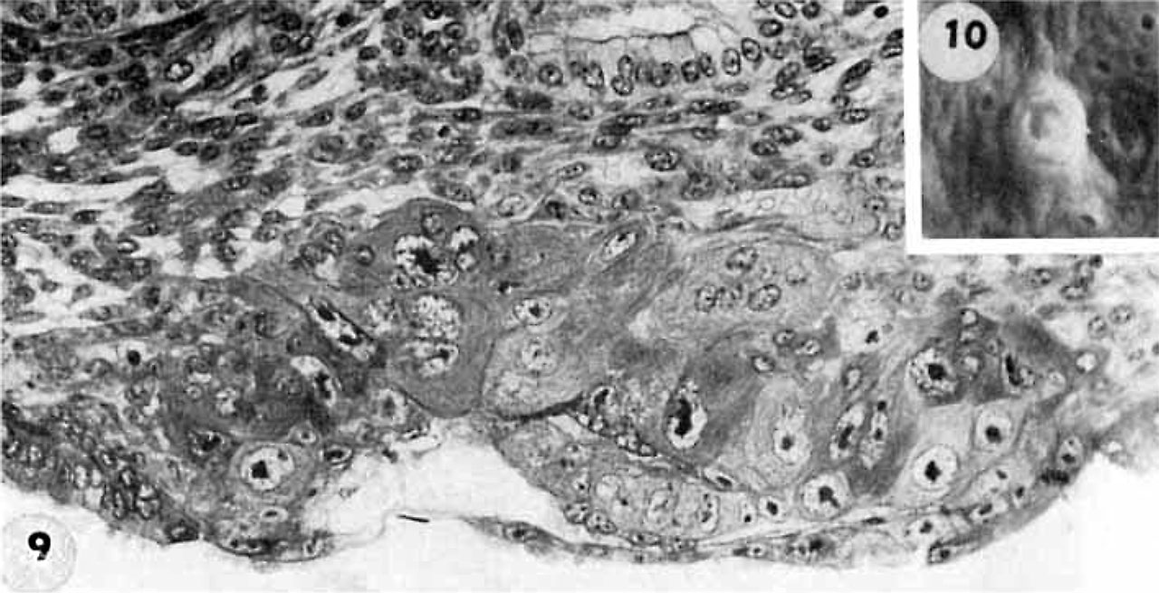
Fig. 9-10. Stage 5a 7.5 days cross-section and surface view
9 A recently implanted ovum of not more than 7.5 days of age, based upon a critical coital date. Note solid trophoblast. composed of paler discrete mononuelear cells and darker less discrete multinucleated cells. The solid disc of tropho~ blast at the embryonic (implantation) pole is continuous with the membranous unaltered blastocyst wall at the abembryonic pole. Note also the amniogenic cells, tiny cleft-like amniotic cavity, large columnar, non-vacuolated ectodermal cells and smaller flattened or polyhedral endodcrmal cells. The ehorionic cavity is irregularly flattened due to collapse of the hlastocyst. Carnegie 8225, Section 20-5-5. X 300.
10 Surface view of the normal 7-day ovum seen in figure 9 photographed under fluid after fixation and partial dehydration. Note embryonic mass within chorionic cavity and the relatively large proportion of the ovum exposed to the uterine lumen. Note prominent gland mouths and surface wrinkling consistent with the 22nd day of the menstrual cycle. Carnegie 8225, Sequence 5. X 22.
- Figure Links: 1 | 2 | 3 | 4 | 5 | 6 | 7 | 8 | 9-10 | 11-12 | 13-14 | 15-16 | 17 | 18-19 | 20 | 21-22 | 23 | 24-25 | 26-27 | 28-29 | 30-31 | 32-33 | 34 | 35 | 36 | 37 | 38 | 39 | 40 | 41 | 42 | 43 | 44 | 45 | 46 | 47 | 48 | 40 | 49 | 50 | 51 | 52 | 53 | 54 | 55 | 56 | 57 | 58 | 59 | 60 | 61 | 62 | 63 | 64 | 65 | 66 | 67 | 68 | 69 | 70 | 71 | 72 | 73 | 74 | 75 | 76 | 77 | 78 | 79 | 80 | 81 | 82 | 83 | 84 | 85 | 86 | 87 | 88 | 89 | 90 | plate 1 | plate 2 | plate 3 | plate 4 | plate 5 | plate 6 | plate 7 | plate 8 | plate 9 | plate 10 | plate 11 | plate 12 | plate 13 | plate 14 | plate 15 | plate 16 | plate 17 | table 1 | table 1 image | table 2 image | table 3 image | table 4 | table 4 image | table 5 | table 5 image | All figures | 1956 Hertig | Embryology History - Arthur Hertig | John Rock | Historic Papers
Reference
Hertig AT. Rock J. and Adams EC. A description of 34 human ova within the first 17 days of development. (1956) Amer. J Anat., 98:435-493.
Cite this page: Hill, M.A. (2024, April 19) Embryology Hertig1956 fig09-10.jpg. Retrieved from https://embryology.med.unsw.edu.au/embryology/index.php/File:Hertig1956_fig09-10.jpg
- © Dr Mark Hill 2024, UNSW Embryology ISBN: 978 0 7334 2609 4 - UNSW CRICOS Provider Code No. 00098G
File history
Click on a date/time to view the file as it appeared at that time.
| Date/Time | Thumbnail | Dimensions | User | Comment | |
|---|---|---|---|---|---|
| current | 19:48, 23 February 2017 | | 1,280 × 655 (166 KB) | Z8600021 (talk | contribs) |
You cannot overwrite this file.
File usage
The following 3 pages use this file: