File:Hertig1945d fig18.jpg
From Embryology
Size of this preview: 397 × 600 pixels. Other resolution: 634 × 958 pixels.
Original file (634 × 958 pixels, file size: 191 KB, MIME type: image/jpeg)
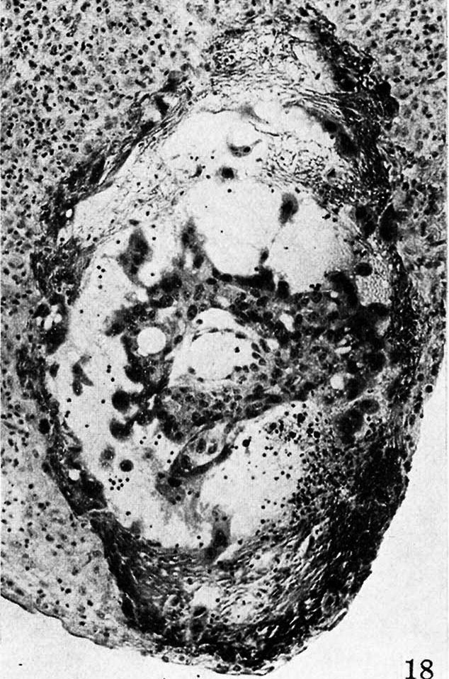
Fig. 18. A mid-cross section of a pathologic, pre-villous ovum of uncertain age but probably 11 to 12 days old
Its embryonic mass and hence the amnion are absent although there is an exioocoelomic membrane and some mesoblastic tissue lining the chorionic cavity.
Carnegie 7771, section 3-4-I. x 100.
Reference
Hertig AT. On the development of the amnion and exocoelomic membrane in the previllous human ovum. (1945) Yale J Biol Med. 18:107-15. PubMed 21007544
Cite this page: Hill, M.A. (2024, April 19) Embryology Hertig1945d fig18.jpg. Retrieved from https://embryology.med.unsw.edu.au/embryology/index.php/File:Hertig1945d_fig18.jpg
- © Dr Mark Hill 2024, UNSW Embryology ISBN: 978 0 7334 2609 4 - UNSW CRICOS Provider Code No. 00098G
File history
Click on a date/time to view the file as it appeared at that time.
| Date/Time | Thumbnail | Dimensions | User | Comment | |
|---|---|---|---|---|---|
| current | 09:10, 8 April 2020 | | 634 × 958 (191 KB) | Z8600021 (talk | contribs) |
You cannot overwrite this file.
File usage
The following page uses this file: