File:Hertig1945d fig03.jpg: Difference between revisions
No edit summary |
mNo edit summary |
||
| (One intermediate revision by the same user not shown) | |||
| Line 1: | Line 1: | ||
==Fig. 3. A section of a. 7.5-day ovum== | |||
The same ovum as fig. 2 but 7 sections removed. The amniotic cavity is less evident because of its smaller size although the amniogenic cells overlying it are more prominent. Carnegie No. {{CE8020}}, section 6-5-2, X 300. | |||
<gallery> | |||
Hertig1945d fig01.jpg|Fig 1 Carnegie No. {{CE8225}} | |||
Hertig1945d fig02.jpg|Fig 2 Carnegie No. {{CE8020}} | |||
Hertig1945d fig03.jpg|Fig 3 Carnegie No. {{CE8020}} | |||
Hertig1945d fig04.jpg|Fig 4 Carnegie No. {{CE8020}} | |||
Hertig1945d fig05.jpg|Fig 5 Carnegie No. {{CE8020}} | |||
</gallery> | |||
===Reference=== | |||
{{Ref-Hertig1945d}} | |||
{{Footer}} | |||
[[Category:Carnegie Embryo 8020]] | |||
Latest revision as of 15:22, 24 October 2017
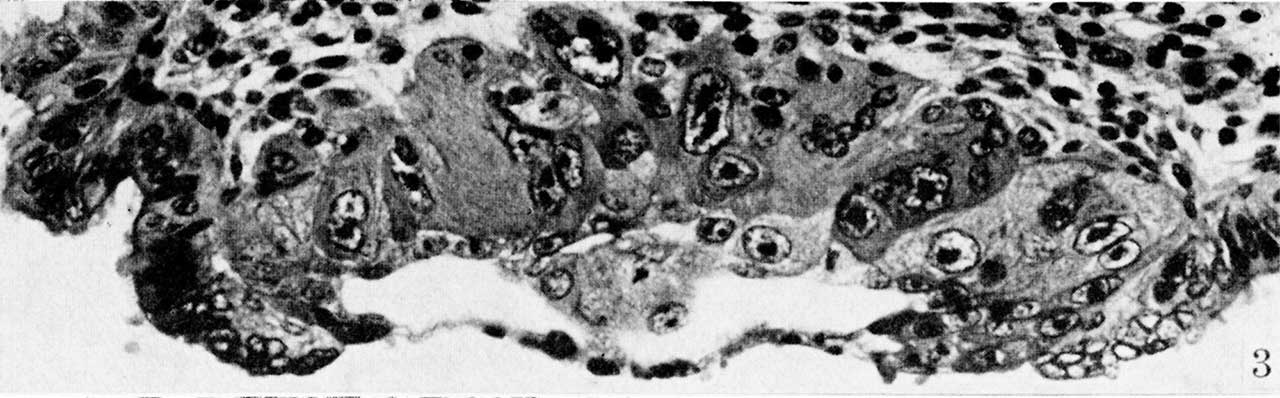
Fig. 3. A section of a. 7.5-day ovum
The same ovum as fig. 2 but 7 sections removed. The amniotic cavity is less evident because of its smaller size although the amniogenic cells overlying it are more prominent. Carnegie No. 8020, section 6-5-2, X 300.
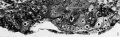
Fig 1 Carnegie No. 8225
Fig 2 Carnegie No. 8020
Fig 3 Carnegie No. 8020
Fig 4 Carnegie No. 8020
Fig 5 Carnegie No. 8020
Reference
Hertig AT. On the development of the amnion and exocoelomic membrane in the previllous human ovum. (1945) Yale J Biol Med. 18:107-15. PubMed 21007544
Cite this page: Hill, M.A. (2024, April 19) Embryology Hertig1945d fig03.jpg. Retrieved from https://embryology.med.unsw.edu.au/embryology/index.php/File:Hertig1945d_fig03.jpg
- © Dr Mark Hill 2024, UNSW Embryology ISBN: 978 0 7334 2609 4 - UNSW CRICOS Provider Code No. 00098G
File history
Click on a date/time to view the file as it appeared at that time.
| Date/Time | Thumbnail | Dimensions | User | Comment | |
|---|---|---|---|---|---|
| current | 15:17, 24 October 2017 | 1,280 × 398 (87 KB) | Z8600021 (talk | contribs) |
You cannot overwrite this file.
File usage
The following 7 pages use this file: