File:Hedgehog signaling pathway.jpg: Difference between revisions
mNo edit summary |
mNo edit summary |
||
| Line 12: | Line 12: | ||
:'''Links:''' [[Developmental Signals - Sonic hedgehog|Sonic hedgehog]] | :'''Links:''' [[Developmental Signals - Sonic hedgehog|Sonic hedgehog]] | ||
{{Factor Links}} | |||
===Reference=== | ===Reference=== | ||
<pubmed>19040769</pubmed>| [http://www.ncbi.nlm.nih.gov/pmc/articles/PMC2614485 PMC2614485] | [http://genomebiology.com/content/9/11/241 Genome Biology] | <pubmed>19040769</pubmed>| [http://www.ncbi.nlm.nih.gov/pmc/articles/PMC2614485 PMC2614485] | [http://genomebiology.com/content/9/11/241 Genome Biology] | ||
| Line 22: | Line 23: | ||
Gb-2011-12-5-222-5.jpg | Gb-2011-12-5-222-5.jpg | ||
{{Footer}} | |||
[[Category:Cartoon]][[Category:Sonic Hedgehog]] | [[Category:Cartoon]][[Category:Sonic Hedgehog]] | ||
Latest revision as of 08:29, 7 October 2015
Hedgehog Signaling Pathway
A simplified Hh signaling pathway, constructed from combined Drosophila and mammalian data.
Hh is targeted to the endoplasmic reticulum by its signal peptide, is palmitoylated at its amino terminus by Rasp/Skinny Hedgehog (Ski), and autoprocessed. Lipidated HhN (M-HhN) is released by Dispatched (Disp) and forms multimers or associates with lipoproteins (LP) in the extracellular environment [32]. A number of molecules can interact with M-HhN and propagate or modulate its trafficking: the Dally-like protein (Dlp), which is modified by the heparan sulfate (HS) polymerases Tout-velu (Ttv), Sister of tout-velu (Sotv), and Brother of tout-velu (Botv), all members of the EXT family; the Hedgehog-interacting protein (Hip); and the Growth-arrest-specific 1 (Gas1) protein. Hip and Gas1 are not present in Drosophila. Megalin (Meg) is most probably involved in the recycling of M-HhN. Ihog is thought to function as co-receptor for M-HhN. M-HhN acts as an antagonistic ligand that represses the function of the receptor Patched (Ptc), a 12-transmembrane protein related to Disp. Binding of M-HhN to Ptc results in internalization. Smoothened (Smo) is a seven-pass membrane receptor, which is key for the transmission of the signal to the nucleus in the Hh pathway. Smo is inhibited by Ptc when not bound by M-HhN. When the inhibitory function of Ptc is released by M-HhN, Smo can translocate to the plasma membrane or - in mammals - to the primary cilium, and active Smo is phosphorylated (red P). Ptc may secrete pro-vitamin D3 or related compounds (D3) to inhibit Smo. Conversely, oxysterols (Oxy) can indirectly activate Smo [52,55]. The Hh pathway downstream of Smo displays some important differences between Drosophila and mammals.
In Drosophila, when Smo is active, the signal passes through a complex comprising the kinesin-like molecule Costal 2 (Cos2), Fused (Fu), Suppressor of fused (Su(fu)) and Cubitus interruptus (Ci), leading to the release of Ci, which can then enter the nucleus to activate transcription. When Smo is inhibited, the Cos2/Fu/Su(fu)/Ci complex remains associated with microtubules, Ci is phosphorylated and is cleaved by Cos2. The Ci fragment now acts as a transcriptional repressor.
In mammals, the targeting of Smo to primary cilia is essential for signal transduction. No obvious equivalents of Cos2 and Fu exist in mammals. Instead, Su(fu) has a more prominent role in inhibiting the pathway. Gli1, Gli2, and Gli3 are the mammalian homologs of Ci; Gli1 and Gli2 activate transcription when Smo is active, whereas Gli3 is processed and becomes a repressor when Smo is inhibited. A number of components in the pathway, in particular downstream of Smo, are not shown in this figure.
- Links: Sonic hedgehog
| Factor Links: AMH | hCG | BMP | sonic hedgehog | bHLH | HOX | FGF | FOX | Hippo | LIM | Nanog | NGF | Nodal | Notch | PAX | retinoic acid | SIX | Slit2/Robo1 | SOX | TBX | TGF-beta | VEGF | WNT | Category:Molecular |
Reference
<pubmed>19040769</pubmed>| PMC2614485 | Genome Biology
Bürglin Genome Biology 2008 9:241 doi:10.1186/gb-2008-9-11-241
Copyright
Authors of Research Articles, Methodologies and Software articles published in Genome Biology are the copyright holders of their articles and have granted to any third party, in advance and in perpetuity, the right to use, reproduce or disseminate the article, according to the BioMed Central copyright and license agreement.
Gb-2011-12-5-222-5.jpg
Cite this page: Hill, M.A. (2024, April 18) Embryology Hedgehog signaling pathway.jpg. Retrieved from https://embryology.med.unsw.edu.au/embryology/index.php/File:Hedgehog_signaling_pathway.jpg
- © Dr Mark Hill 2024, UNSW Embryology ISBN: 978 0 7334 2609 4 - UNSW CRICOS Provider Code No. 00098G
File history
Click on a date/time to view the file as it appeared at that time.
| Date/Time | Thumbnail | Dimensions | User | Comment | |
|---|---|---|---|---|---|
| current | 07:47, 13 June 2012 | 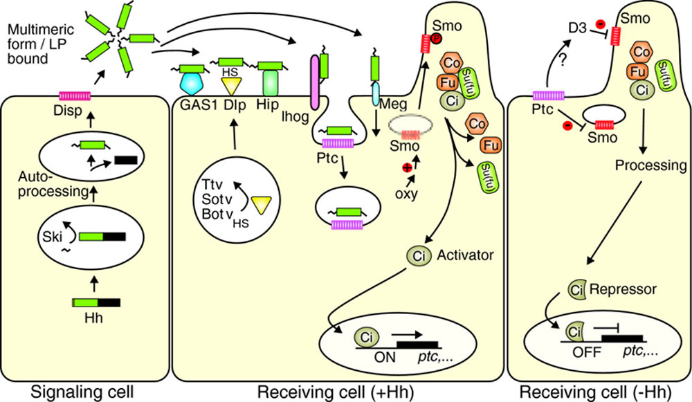 | 1,000 × 581 (102 KB) | Z8600021 (talk | contribs) | ==Hedgehog Signaling Pathway== A simplified Hh signaling pathway, constructed from combined Drosophila and mammalian data. Hh is targeted to the endoplasmic reticulum by its signal peptide, is palmitoylated at its amino terminus by Rasp/Skinny Hedgehog |
You cannot overwrite this file.