File:Heart chicken embryo stage 12.jpg
From Embryology

Size of this preview: 507 × 599 pixels. Other resolution: 1,000 × 1,182 pixels.
Original file (1,000 × 1,182 pixels, file size: 221 KB, MIME type: image/jpeg)
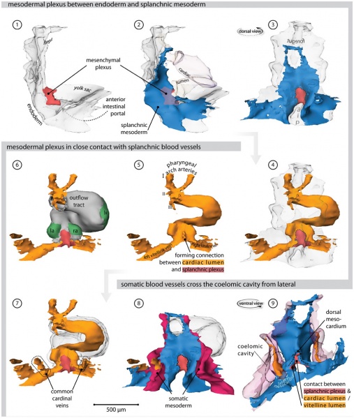
Chicken Heart (stage 12 embryo)
- Right lateral view of the endoderm (transparent). Overlying the endoderm of the anterior intestinal portal is a mesenchymal plexus (red).
- Overlying this mesenchymal plexus is the splanchnic mesoderm (blue). The lumen of the heart tube is transparent.
- Dorsal view, showing how the vascular plexus is wedged between the endoderm and the splanchnic mesoderm.
- The cardiovascular lumen is shown in orange; the forming vitelline veins are continuous with the vessels that cover the yolk sac.
- Cardiac lumen protrudes into dorsal mesocardium, where contact with splanchnic plexus is being established.
- Bilaterally flanking the dorsal mesocardium are the emerging atria, as distinguished by expression of Connexin40 (green).
- Hooking into the vitelline veins from dorsolateral are the common cardinal veins.
- These common cardinal veins reside in somatic mesoderm (red).
- Ventral view of the splanchnic and somatic mesoderm. Separating these tissues is the coelomic cavity. They contact at the so-called lateral mesocardium (light blue).
Abbreviations
- I, II, III - first, second & third pharyngeal arch artery
- aip - anterior intestinal portal
- la - left atrium
- ra - right atrium
- lv - left ventricle
Supplemental File S1 for an interactive version of this reconstruction.
- Links: Image - Heart 3D reconstruction | Image - Stage 12 Heart | Image - Stage 16 Heart | Image - Stage 21 Heart | Image - Stage 25 Heart | Hamburger Hamilton Stages | Cardiovascular System Development | Chicken Development
Original image name: Figure 3. Journal.pone.0022055.g003.jpg http://www.ncbi.nlm.nih.gov/pmc/articles/PMC3133620/figure/pone-0022055-g003/
Reference
<pubmed>21779373</pubmed>| PLoS One
© 2011 van den Berg, Moorman. This is an open-access article distributed under the terms of the Creative Commons Attribution License, which permits unrestricted use, distribution, and reproduction in any medium, provided the original author and source are credited.
File history
Click on a date/time to view the file as it appeared at that time.
| Date/Time | Thumbnail | Dimensions | User | Comment | |
|---|---|---|---|---|---|
| current | 09:08, 27 August 2011 |  | 1,000 × 1,182 (221 KB) | S8600021 (talk | contribs) | ==Chicken Heart (stage 12 embryo)== # Right lateral view of the endoderm (transparent). Overlying the endoderm of the anterior intestinal portal is a mesenchymal plexus (red). # Overlying this mesenchymal plexus is the splanchnic mesoderm (blue). The lum |
You cannot overwrite this file.
File usage
The following 2 pages use this file: