File:Hearing cartoon.jpg: Difference between revisions
From Embryology
No edit summary |
|||
| (One intermediate revision by one other user not shown) | |||
| Line 1: | Line 1: | ||
==Adult Hearing Components== | |||
Cartoon showing the 3 main divisions of the ear. | Cartoon showing the 3 main divisions of the ear. | ||
Image | '''Related images:''' [[:File:Adult hearing embryonic origins.jpg|Image - Adult hearing embryonic origins]] | [[:File:Hearing_cartoon.jpg|Image - without embryology]] | ||
{{Hearing Links}} | |||
{{Footer}} | |||
[[Category:Senses]] [[Category:Hearing]] [[Category:Cartoon]] | [[Category:Senses]] [[Category:Hearing]] [[Category:Cartoon]] | ||
Latest revision as of 08:05, 7 April 2016
Adult Hearing Components
Cartoon showing the 3 main divisions of the ear.
Related images: Image - Adult hearing embryonic origins | Image - without embryology
Cite this page: Hill, M.A. (2024, April 18) Embryology Hearing cartoon.jpg. Retrieved from https://embryology.med.unsw.edu.au/embryology/index.php/File:Hearing_cartoon.jpg
- © Dr Mark Hill 2024, UNSW Embryology ISBN: 978 0 7334 2609 4 - UNSW CRICOS Provider Code No. 00098G
File history
Click on a date/time to view the file as it appeared at that time.
| Date/Time | Thumbnail | Dimensions | User | Comment | |
|---|---|---|---|---|---|
| current | 20:44, 13 May 2009 | 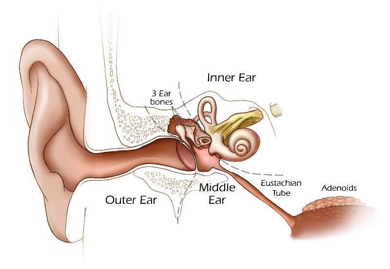 | 800 × 553 (43 KB) | MarkHill (talk | contribs) | Cartoon showing the 3 main divisions of the ear. Image source: UNSW Embryology, modified from NIH image. |
You cannot overwrite this file.
File usage
The following 21 pages use this file:
- 2009 BGD-B Lecture Face and Ear
- 2009 Lecture 17
- 2010 Lecture 17
- 2011 Lab 10
- ANAT2341 Lab 6 - Early Embryo
- BGDB Face and Ear - Early Embryo
- Human System Development
- K12 - Communication
- K12 Professional Development 2014
- Lecture - Sensory Development
- Sensory - Hearing Abnormalities
- Sensory - Hearing and Balance Development
- Sensory System - Abnormalities
- Sensory System Development
- Talk:2009 Lecture 17
- Talk:ANAT2341 Lab 10
- Talk:Hearing - Outer Ear Development
- Talk:Sensory System Development
- Category:Inner Ear
- Category:Middle Ear
- Category:Outer Ear