File:Hearing-vestibular sac abnormality.jpg
Hearing-vestibular_sac_abnormality.jpg (432 × 493 pixels, file size: 45 KB, MIME type: image/jpeg)
Vestibular Sac Abnormality
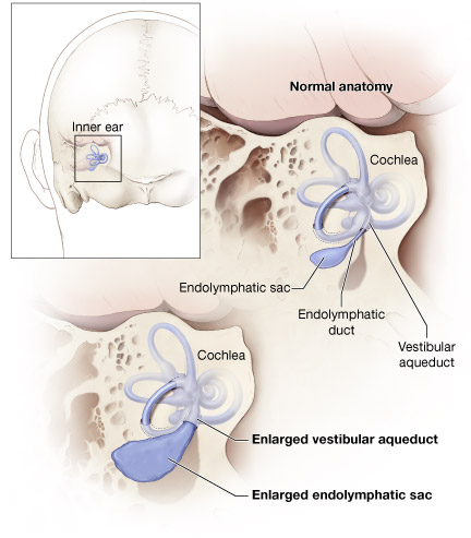
Diagram of normal inner ear and enlarged vestibular aqueduct, showing the cochlea, endolymphatic sac, endolymphatic duct, vestibular aqueduct, enlarged vestibular aqueduct, and enlarged endolymphatic sac.
Original file name: VestAque_lg.jpg http://www.nidcd.nih.gov/health/hearing/vestAque.htm
NIDCD web site [Copyright Policy]
Unless otherwise stated, the information on this site is not copyrighted and is in the public domain. It is free for the public to use, copy, and distribute. You may encounter documents that were sponsored along with private companies or other organizations. Those documents will have statements that protect them under U.S. and foreign copyright laws.
Related Developmental Information
The information below is modified from article abstracts.
Postnatal development of the vestibular aqueduct and endolymphatic sac
- "The purpose of this study was to gain basic information about the postnatal development of the vestibular aqueduct (VA) and the endolymphatic sac (ES). For this study, serial horizontal sections of 31 normal temporal bones of individuals whose ages ranged from 0 to 13 years were used. Medial view graphic reconstruction of the VA and rugose portion (RP) of the ES was performed in every case for analysis of the VA and RP. The findings of this study revealed the following new information about the postnatal development of the VA and ES.
- The VA and RP undergo significant growth postnatally up to age 3 years.
- In the newborn, individual variations in the VA and RP already exist and at age 3 years significantly wide individual variations which can be classified into three groups (hypoplastic, normoplastic, hyperplastic) may be recognized.
- Hypoplastic VAs are of two types: one is fairly elongated and tubelike while the other is short and funnel-shaped. The tubelike VA seems to be the prenatal form.
- The changes that occur with development postnatally in the area of the VA are more closely related to the changes that occur in the length of the external aperture of the VA than they are to the changes that occur in the length of the VA.
- Development of the area of the VA is closely correlated with development of the area of the RP."[1]
Morphological analysis of the vestibular aqueduct by computerized tomography images
- "The morphology of the vestibular aqueduct may be funnel-shaped, filiform or tubular and the respective proportions were found to be at 44%, 33% and 22% in children and 21.7%, 53.3% and 25% in adults. The morphometric data showed to be of 4.86 mm(2) of area, 2.24 mm of the external opening, 4.73 mm of length and 11.88 mm of the distance from the vestibular aqueduct to the internal acoustic meatus, in children, and in adults it was of 4.93 mm(2), 2.09 mm, 4.44 mm, and 11.35 mm, respectively."[2]
References
Related References
File history
Click on a date/time to view the file as it appeared at that time.
| Date/Time | Thumbnail | Dimensions | User | Comment | |
|---|---|---|---|---|---|
| current | 08:05, 27 September 2010 |  | 432 × 493 (45 KB) | S8600021 (talk | contribs) | ==Vestibular Sac Abnormality== Diagram of normal inner ear and enlarged vestibular aqueduct, showing the cochlea, endolymphatic sac, endolymphatic duct, vestibular aqueduct, enlarged vestibular aqueduct, and enlarged endolymphatic sac. Original file name |
You cannot overwrite this file.
File usage
The following 3 pages use this file:
