File:HansonAnson1962 fig02.jpg
Original file (1,280 × 601 pixels, file size: 233 KB, MIME type: image/jpeg)
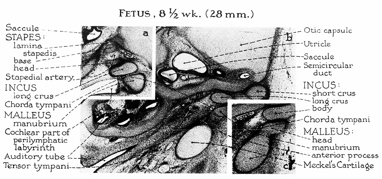
Fig. 2. 8.5 week 28 mm
a, At this level the ‘‘stapes”’ (still shaped like a ring rather than a stirrup) presses against the lateral wall of the cartilaginous otic capsule to form the lamina stapedis. The long crus of the incus approximates the future capital part of the stapes but is separated from the manubrium by the chorda tympani. The loose mesenchyme is being invaded by the first parhyngeal pouch, which eventually will line the tympanic cavity and envelop the ossicles. The anterior process of malleus appears for the first time as a spicule of membrane bone situated medialto Meckel’s cartilage and anterior to the manubrium. [tis the only portion of the ossicles which is not formed in cartilage.
b, The ossicles are fully formed in cartilage between the eighth and ninth{weeks of fetal life. A dense zone of mesenchymal cells renders the two ossicles distinguishable from each other.
c, Meckel’s cartilage is broadly continuous with the head of the malleus.
Reference
Hanson JR. and Anson BJ. Development of the malleus of the human ear; Illustrated in atlas series. (1962) Q Bull Northwest Univ Med Sch. 36(2): 119–137. PMID: 13904457.
Cite this page: Hill, M.A. (2024, April 16) Embryology HansonAnson1962 fig02.jpg. Retrieved from https://embryology.med.unsw.edu.au/embryology/index.php/File:HansonAnson1962_fig02.jpg
- © Dr Mark Hill 2024, UNSW Embryology ISBN: 978 0 7334 2609 4 - UNSW CRICOS Provider Code No. 00098G
File history
Click on a date/time to view the file as it appeared at that time.
| Date/Time | Thumbnail | Dimensions | User | Comment | |
|---|---|---|---|---|---|
| current | 10:17, 7 January 2019 | | 1,280 × 601 (233 KB) | Z8600021 (talk | contribs) | |
| 10:13, 7 January 2019 |  | 1,962 × 1,265 (513 KB) | Z8600021 (talk | contribs) |
You cannot overwrite this file.
File usage
The following 2 pages use this file: