File:HamiltonBoyd1960 plate06.jpg: Difference between revisions
mNo edit summary |
mNo edit summary |
||
| Line 8: | Line 8: | ||
'''Fig. 18.''' Photomicrograph ( x 270) of a section (H. & E.) through a uterine gland near the implantation site of an 18-day embryo (Gar). The lumen of the gland possesses maternal red blood corpuscles and two masses of multinucleated syncytium. | '''Fig. 18.''' Photomicrograph ( x 270) of a section (H. & E.) through a uterine gland near the implantation site of an 18-day embryo (Gar). The lumen of the gland possesses maternal red blood corpuscles and two masses of multinucleated syncytium. | ||
{{HamiltonBoyd1960 plates footer}} | |||
Latest revision as of 13:04, 6 August 2020
Plate 6
Fig. 15. Photomicrograph (x 100) of part of a section (H. & E.) through the foetal-maternal junction of a 14-day embryo (Missen). The chorionic villi (to the left of the illustration) possess mesodermal cores but as yet no definitive blood vessels are present. The villi possess a covering of definitive syncytium overlying Langhan’s layer. At the tips of some of the villi cytotrophoblastic columns, by proliferation, are contributing to the trophoblastic shell. On the maternal side of this shell a number of persisting masses of primitive syncytium can be identified. In the intervillous space there are maternal blood vessels and at about the centre of the figure an isolated syncytial sprout in the intervillous space; maternal blood cells are also present in the intervillous space.
Fig. 16. Photomicrograph ( x 150) of part of a section (H. & E.) through the tips of the chorionic villi (above) and of the adjacent endometrium (below) in a 16-day human embryo. Cytotrophoblastic columns at the tips of the two central villi have proliferated and fused to form part of the trophoblastic shell. Definitive syncytium can be identified on the surface of the villi and primitive syncytium is related to the shell. Maternal blood vessels and a syncytial sprout. can be identified in the intervillous space.
Fig. 17. Photomicrograph ( x 200) of a section (H. & E.) through the peripheral syncytium of an 18-day embryo (Gar). The syncytium shows extensive vacuolation and a maternal red blood corpuscle is present in one of the vacuoles. At another point several such red blood corpuscles are in the process of coming to be included within the syncytium.
Fig. 18. Photomicrograph ( x 270) of a section (H. & E.) through a uterine gland near the implantation site of an 18-day embryo (Gar). The lumen of the gland possesses maternal red blood corpuscles and two masses of multinucleated syncytium.
Plates: 1 | 2 | 3 | 4 | 5 | 6 | 7 | 8 | 9 | 10 | 11 | 12 | 13
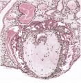
Plate 1
Plate 2
Plate 3
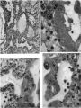
Plate 4
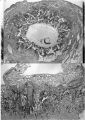
Plate 5
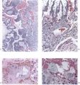
Plate 6
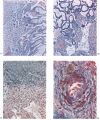
Plate 7
Plate 8
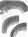
Plate 9
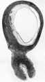
Plate 10
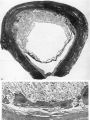
Plate 11
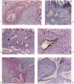
Plate 12
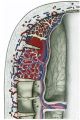
Plate 13


Reference
Hamilton WJ. and Boyd JD. Development of the human placenta in the first three months of gestation. (1960) J Anat. 94(3): 297-328. PMID14399291 | PDF
Cite this page: Hill, M.A. (2024, April 25) Embryology HamiltonBoyd1960 plate06.jpg. Retrieved from https://embryology.med.unsw.edu.au/embryology/index.php/File:HamiltonBoyd1960_plate06.jpg
- © Dr Mark Hill 2024, UNSW Embryology ISBN: 978 0 7334 2609 4 - UNSW CRICOS Provider Code No. 00098G
File history
Click on a date/time to view the file as it appeared at that time.
| Date/Time | Thumbnail | Dimensions | User | Comment | |
|---|---|---|---|---|---|
| current | 12:40, 6 August 2020 |  | 1,280 × 1,354 (444 KB) | Z8600021 (talk | contribs) | crop, adjust size |
| 12:37, 6 August 2020 |  | 1,030 × 1,429 (339 KB) | Z8600021 (talk | contribs) |
You cannot overwrite this file.
File usage
The following 12 pages use this file:
- Paper - Development of the human placenta in the first three months of gestation (1960)
- File:HamiltonBoyd1960 fig02.jpg
- File:HamiltonBoyd1960 fig03.jpg
- File:HamiltonBoyd1960 fig04.jpg
- File:HamiltonBoyd1960 fig05.jpg
- File:HamiltonBoyd1960 fig06.jpg
- File:HamiltonBoyd1960 fig07.jpg
- File:HamiltonBoyd1960 fig08.jpg
- File:HamiltonBoyd1960 plate02.jpg
- File:HamiltonBoyd1960 plate03.jpg
- File:HamiltonBoyd1960 plate13.jpg
- Template:HamiltonBoyd1960 plates