File:HamiltonBoyd1960 plate05.jpg: Difference between revisions
(Z8600021 uploaded a new version of File:HamiltonBoyd1960 plate05.jpg) |
mNo edit summary |
||
| Line 3: | Line 3: | ||
'''Fig. 14.''' Photomicrograph ( x 10) of a section of the implantation site of a 28-somite human embryo (Camb. H. 710). Villi which cover the whole of the chorionic sac are well established, and possess numerous foetal vessels. There is no indication of a marginal sinus in the intervillous space ; there is, however, venous drainage from the left margin of this space. The decidua basalis is thick and contains many dilated glands; several of the central ones show haemorrhage into their lumina. The effect of expansion of the chorionic sac on the uterine glands is particularly well shown on the right of the illustration where their necks are constricted and stretched, thus damming back the products of secretion. Veins and spiral arteries can be identified at intervals. in the decidua basalis. | '''Fig. 14.''' Photomicrograph ( x 10) of a section of the implantation site of a 28-somite human embryo (Camb. H. 710). Villi which cover the whole of the chorionic sac are well established, and possess numerous foetal vessels. There is no indication of a marginal sinus in the intervillous space ; there is, however, venous drainage from the left margin of this space. The decidua basalis is thick and contains many dilated glands; several of the central ones show haemorrhage into their lumina. The effect of expansion of the chorionic sac on the uterine glands is particularly well shown on the right of the illustration where their necks are constricted and stretched, thus damming back the products of secretion. Veins and spiral arteries can be identified at intervals. in the decidua basalis. | ||
{{HamiltonBoyd1960 plates footer}} | |||
Latest revision as of 13:04, 6 August 2020
Plate 5
Fig. 13. Photomicrograph ( x 45) of a section of the implantation site of a 14-day embryo (Missen). Chorionic villi with mesodermal cores are now well established round the whole of the chorionic sac. There is an extensive intervillous space which, in this section, communicates at two points with a large venous sinusoid (S.). Details of the differentiation of the trophoblast in this specimen are shown in PI. 6, fig. 15.
Fig. 14. Photomicrograph ( x 10) of a section of the implantation site of a 28-somite human embryo (Camb. H. 710). Villi which cover the whole of the chorionic sac are well established, and possess numerous foetal vessels. There is no indication of a marginal sinus in the intervillous space ; there is, however, venous drainage from the left margin of this space. The decidua basalis is thick and contains many dilated glands; several of the central ones show haemorrhage into their lumina. The effect of expansion of the chorionic sac on the uterine glands is particularly well shown on the right of the illustration where their necks are constricted and stretched, thus damming back the products of secretion. Veins and spiral arteries can be identified at intervals. in the decidua basalis.
Plates: 1 | 2 | 3 | 4 | 5 | 6 | 7 | 8 | 9 | 10 | 11 | 12 | 13
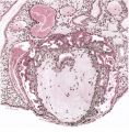
Plate 1
Plate 2
Plate 3
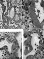
Plate 4

Plate 5
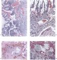
Plate 6
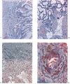
Plate 7
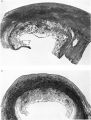
Plate 8
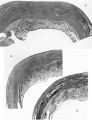
Plate 9
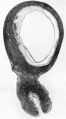
Plate 10
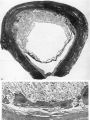
Plate 11
Plate 12
Plate 13


Reference
Hamilton WJ. and Boyd JD. Development of the human placenta in the first three months of gestation. (1960) J Anat. 94(3): 297-328. PMID14399291 | PDF
Cite this page: Hill, M.A. (2024, April 25) Embryology HamiltonBoyd1960 plate05.jpg. Retrieved from https://embryology.med.unsw.edu.au/embryology/index.php/File:HamiltonBoyd1960_plate05.jpg
- © Dr Mark Hill 2024, UNSW Embryology ISBN: 978 0 7334 2609 4 - UNSW CRICOS Provider Code No. 00098G
File history
Click on a date/time to view the file as it appeared at that time.
| Date/Time | Thumbnail | Dimensions | User | Comment | |
|---|---|---|---|---|---|
| current | 12:39, 6 August 2020 | 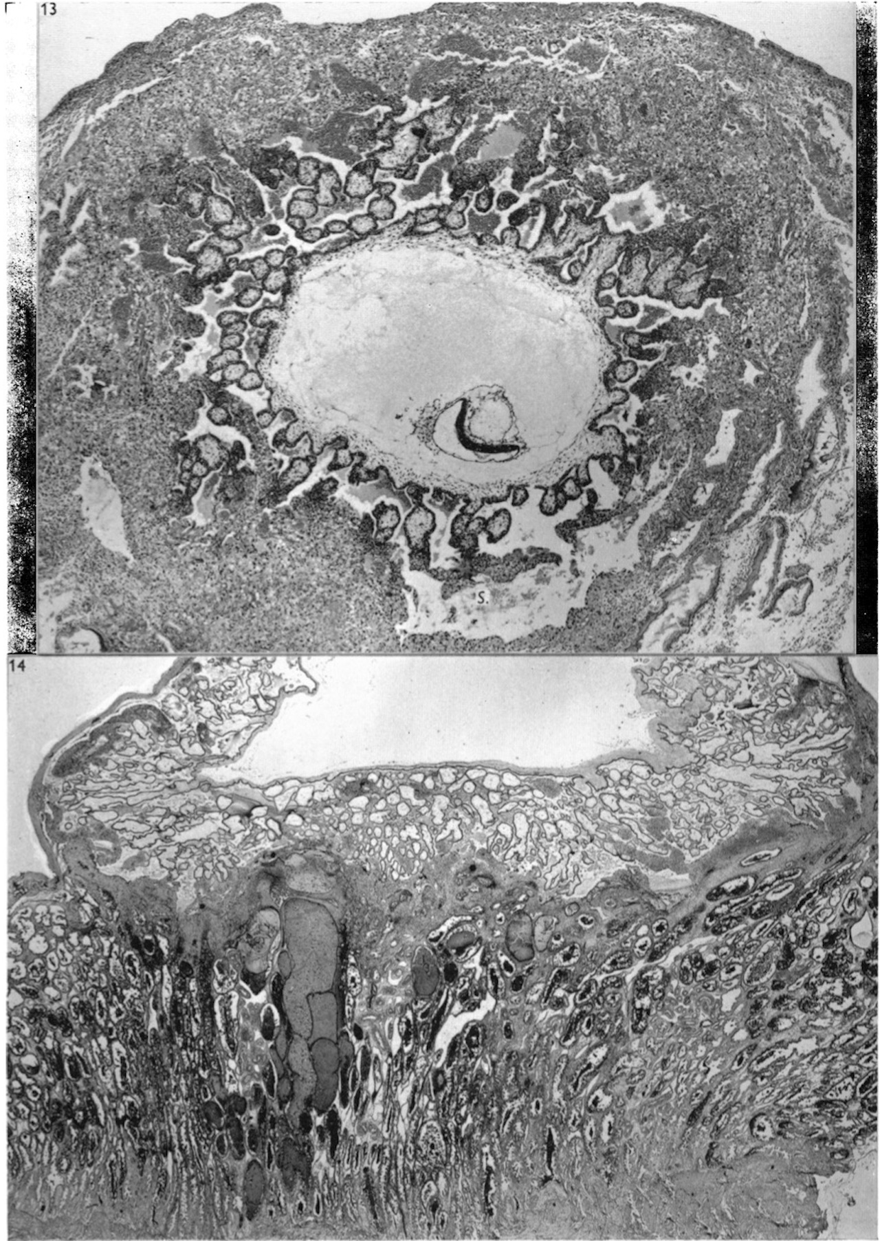 | 1,280 × 1,805 (790 KB) | Z8600021 (talk | contribs) | crop, adjust size |
| 12:37, 6 August 2020 |  | 1,030 × 1,429 (542 KB) | Z8600021 (talk | contribs) |
You cannot overwrite this file.
File usage
The following 12 pages use this file:
- Paper - Development of the human placenta in the first three months of gestation (1960)
- File:HamiltonBoyd1960 fig02.jpg
- File:HamiltonBoyd1960 fig03.jpg
- File:HamiltonBoyd1960 fig04.jpg
- File:HamiltonBoyd1960 fig05.jpg
- File:HamiltonBoyd1960 fig06.jpg
- File:HamiltonBoyd1960 fig07.jpg
- File:HamiltonBoyd1960 fig08.jpg
- File:HamiltonBoyd1960 plate02.jpg
- File:HamiltonBoyd1960 plate03.jpg
- File:HamiltonBoyd1960 plate13.jpg
- Template:HamiltonBoyd1960 plates