File:HD patients.jpg: Difference between revisions
(Fig 1. Phenotypically identical twins (A) and skull computed tomography showing head of caudate nucleus atrophy (B and C) Patients were female, 24 years-old when first examined, and phenotypically identical (Fig 1A). The onset of their disease, as report) |
No edit summary |
||
| (One intermediate revision by one other user not shown) | |||
| Line 2: | Line 2: | ||
Patients were female, 24 years-old when first examined, and phenotypically identical (Fig 1A). The onset of their disease, as reported by relatives, was at 17 and 20 years of age, the initial manifestations being balance and gait impairment with a progressive course. Neurologic examination showed bradykinesia, rigidity with cogwheel phenomenon, choreic movements of trunk and limbs (mild degree), dysarthria, hyperactive tendon reflexes and flexor plantar responses. The patient with earlier onset of the disease was more severely affected. Neurologic examination was repeated approximately six months and one year after the initial examination, and both patients worsened on each successive examination. | Patients were female, 24 years-old when first examined, and phenotypically identical (Fig 1A). The onset of their disease, as reported by relatives, was at 17 and 20 years of age, the initial manifestations being balance and gait impairment with a progressive course. Neurologic examination showed bradykinesia, rigidity with cogwheel phenomenon, choreic movements of trunk and limbs (mild degree), dysarthria, hyperactive tendon reflexes and flexor plantar responses. The patient with earlier onset of the disease was more severely affected. Neurologic examination was repeated approximately six months and one year after the initial examination, and both patients worsened on each successive examination. | ||
<ref><pubmed>10751926</pubmed></ref> | |||
==Reference== | ==Reference== | ||
<references/> | |||
Creative Commons License All the contents of this journal, except where otherwise noted, is licensed under a Creative Commons Attribution License | Creative Commons License All the contents of this journal, except where otherwise noted, is licensed under a Creative Commons Attribution License | ||
===Assessment=== | |||
+ Figure relavent to group project. | |||
+ Copyright, citation and student disclaimer included. | |||
- Only original figure legend with file. Requires further explanation for peer teaching component. | |||
{{Template:2011 Student Image}} | {{Template:2011 Student Image}} | ||
Latest revision as of 12:15, 28 October 2011
Fig 1. Phenotypically identical twins (A) and skull computed tomography showing head of caudate nucleus atrophy (B and C)
Patients were female, 24 years-old when first examined, and phenotypically identical (Fig 1A). The onset of their disease, as reported by relatives, was at 17 and 20 years of age, the initial manifestations being balance and gait impairment with a progressive course. Neurologic examination showed bradykinesia, rigidity with cogwheel phenomenon, choreic movements of trunk and limbs (mild degree), dysarthria, hyperactive tendon reflexes and flexor plantar responses. The patient with earlier onset of the disease was more severely affected. Neurologic examination was repeated approximately six months and one year after the initial examination, and both patients worsened on each successive examination.
Reference
- ↑ <pubmed>10751926</pubmed>
Creative Commons License All the contents of this journal, except where otherwise noted, is licensed under a Creative Commons Attribution License
Assessment
+ Figure relavent to group project. + Copyright, citation and student disclaimer included. - Only original figure legend with file. Requires further explanation for peer teaching component.
- Note - This image was originally uploaded as part of a student project and may contain inaccuracies in either description or acknowledgements. Students have been advised in writing concerning the reuse of content and may accidentally have misunderstood the original terms of use. If image reuse on this non-commercial educational site infringes your existing copyright, please contact the site editor for immediate removal.
Cite this page: Hill, M.A. (2024, April 19) Embryology HD patients.jpg. Retrieved from https://embryology.med.unsw.edu.au/embryology/index.php/File:HD_patients.jpg
- © Dr Mark Hill 2024, UNSW Embryology ISBN: 978 0 7334 2609 4 - UNSW CRICOS Provider Code No. 00098G
File history
Click on a date/time to view the file as it appeared at that time.
| Date/Time | Thumbnail | Dimensions | User | Comment | |
|---|---|---|---|---|---|
| current | 16:54, 1 October 2011 | 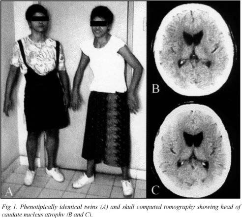 | 492 × 442 (52 KB) | Z3290379 (talk | contribs) | Fig 1. Phenotypically identical twins (A) and skull computed tomography showing head of caudate nucleus atrophy (B and C) Patients were female, 24 years-old when first examined, and phenotypically identical (Fig 1A). The onset of their disease, as report |
You cannot overwrite this file.
File usage
The following 2 pages use this file: