File:Gray1174.jpg
Original file (782 × 800 pixels, file size: 165 KB, MIME type: image/jpeg)
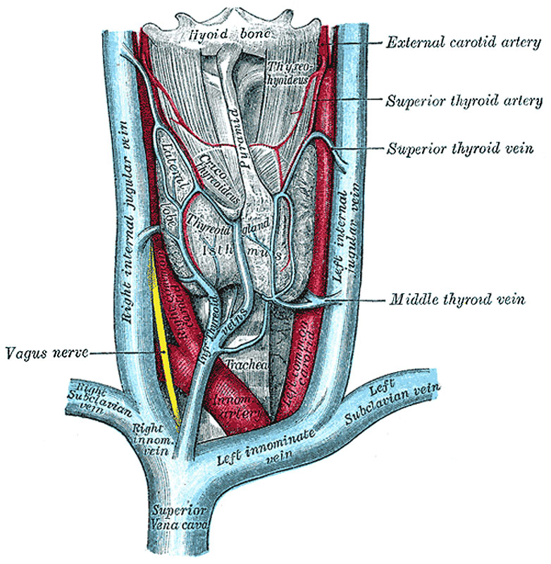
The thyroid gland and its relations
The Thyroid Gland (Glandula Thyreiodea; Thyroid Body) (Fig. 1174).—The thyroid gland is a highly vascular organ, situated at the front and sides of the neck; it consists of right and left lobes connected across the middle line by a narrow portion, the isthmus. Its weight is somewhat variable, but is usually about 30 grams. It is slightly heavier in the female, in whom it becomes enlarged during menstruation and pregnancy.
The lobes (lobuli gl. thyreoideæ) are conical in shape, the apex of each being directed upward and lateralward as far as the junction of the middle with the lower third of the thyroid cartilage; the base looks downward, and is on a level with the fifth or sixth tracheal ring. Each lobe is about 5 cm. long; its greatest width is about 3 cm., and its thickness about 2 cm. The lateral or superficial surface is convex, and covered by the skin, the superficial and deep fasciæ, the Sternocleidomastoideus, the superior belly of the Omohyoideus, the Sternohyoideus and Sternothyreoideus, and beneath the last muscle by the pretracheal layer of the deep fascia, which forms a capsule for the gland. The deep or medial surface is moulded over the underlying structures, viz., the thyroid and cricoid cartilages, the trachea, the Constrictor pharyngis inferior and posterior part of the Cricothyreoideus, the esophagus (particularly on the left side of the neck), the superior and inferior thyroid arteries, and the recurrent nerves. The anterior border is thin, and inclines obliquely from above downward toward the middle line of the neck, while the posterior border is thick and overlaps the common carotid artery, and, as a rule, the parathyroids.
The isthmus (isthmus gl. thyreoidea) connects together the lower thirds of the lobes; it measures about 1.25 cm. in breadth, and the same in depth, and usually covers the second and third rings of the trachea. Its situation and size present, however, many variations. In the middle line of the neck it is covered by the skin and fascia, and close to the middle line, on either side, by the Sternothyreoideus. Across its upper border runs an anastomotic branch uniting the two superior thyroid arteries; at its lower border are the inferior thyroid veins. Sometimes the isthmus is altogether wanting.
A third lobe, of conical shape, called the pyramidal lobe, frequently arises from the upper part of the isthmus, or from the adjacent portion of either lobe, but most commonly the left, and ascends as far as the hyoid bone. It is occasionally quite detached, or may be divided into two or more parts.
A fibrous or muscular band is sometimes found attached, above, to the body of the hyoid bone, and below to the isthmus of the gland, or its pyramidal lobe. When muscular, it is termed the Levator glandulæ thyreoideæ.
Small detached portions of thyroid tissue are sometimes found in the vicinity of the lateral lobes or above the isthmus; they are called accessory thyroid glands (glandulæ thyreoideæ accessoriæ).
- Links: Bold textThyroid Development | Endocrine Development
- Gray's Images: Development | Lymphatic | Neural | Vision | Hearing | Somatosensory | Integumentary | Respiratory | Gastrointestinal | Urogenital | Endocrine | Surface Anatomy | iBook | Historic Disclaimer
| Historic Disclaimer - information about historic embryology pages |
|---|
 |
| iBook - Gray's Embryology | |
|---|---|

|
|
Reference
Gray H. Anatomy of the human body. (1918) Philadelphia: Lea & Febiger.
Cite this page: Hill, M.A. (2024, April 16) Embryology Gray1174.jpg. Retrieved from https://embryology.med.unsw.edu.au/embryology/index.php/File:Gray1174.jpg
- © Dr Mark Hill 2024, UNSW Embryology ISBN: 978 0 7334 2609 4 - UNSW CRICOS Provider Code No. 00098G
File history
Click on a date/time to view the file as it appeared at that time.
| Date/Time | Thumbnail | Dimensions | User | Comment | |
|---|---|---|---|---|---|
| current | 12:13, 25 February 2013 | | 782 × 800 (165 KB) | Z8600021 (talk | contribs) | ==The thyroid gland and its relations== {{Gray Anatomy}} Category:Human Category:Thyroid Category:Cardiovascular |
You cannot overwrite this file.
File usage
The following 2 pages use this file: