File:Gray1137.jpg
Gray1137.jpg (600 × 502 pixels, file size: 64 KB, MIME type: image/jpeg)
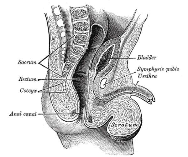
The Bladder in the Child
Sagittal section through the pelvis of a newly born male child.
In the newborn child the internal urethral orifice is at the level of the upper border of the symphysis pubis; the bladder therefore lies relatively at a much higher level in the infant than in the adult. Its anterior surface “is in contact with about the lower two-thirds of that part of the abdominal wall which lies between the symphysis pubis and the umbilicus” (Symington 177). Its fundus is clothed with peritoneum as far as the level of the internal orifice of the urethra. Although the bladder of the infant is usually described as an abdominal organ, Symington has pointed out that only about one-half of it lies above the plane of the superior aperture of the pelvis. Disse maintains that the internal urethral orifice sinks rapidly during the first years, and then more slowly until the ninth year, after which it remains sta when it again slowly descends and reaches its adult position.
- Gray's Images: Development | Lymphatic | Neural | Vision | Hearing | Somatosensory | Integumentary | Respiratory | Gastrointestinal | Urogenital | Endocrine | Surface Anatomy | iBook | Historic Disclaimer
| Historic Disclaimer - information about historic embryology pages |
|---|
 |
| iBook - Gray's Embryology | |
|---|---|

|
|
Reference
Gray H. Anatomy of the human body. (1918) Philadelphia: Lea & Febiger.
Cite this page: Hill, M.A. (2024, April 18) Embryology Gray1137.jpg. Retrieved from https://embryology.med.unsw.edu.au/embryology/index.php/File:Gray1137.jpg
- © Dr Mark Hill 2024, UNSW Embryology ISBN: 978 0 7334 2609 4 - UNSW CRICOS Provider Code No. 00098G
File history
Click on a date/time to view the file as it appeared at that time.
| Date/Time | Thumbnail | Dimensions | User | Comment | |
|---|---|---|---|---|---|
| current | 22:22, 28 October 2010 | | 600 × 502 (64 KB) | S8600021 (talk | contribs) |
You cannot overwrite this file.
File usage
The following 3 pages use this file: