File:Gray1128.jpg: Difference between revisions
No edit summary |
m (→Minute Anatomy) |
||
| (3 intermediate revisions by the same user not shown) | |||
| Line 1: | Line 1: | ||
==Renal - Adult Nephron Structure== | ==Renal - Adult Nephron Structure== | ||
Scheme of renal tubule and its vascular supply. | |||
===Minute Anatomy=== | ===Minute Anatomy=== | ||
The renal tubules (Fig. | The renal tubules ([[:File:Gray1128.jpg|Fig. 1128]]), of which the kidney is for the most part made up, commence in the cortical substance, and after pursuing a very circuitous course through the cortical and medullary substances, finally end at the apices of the renal pyramids by open mouths, so that the fluid which they contain is emptied, through the calyces, into the pelvis of the kidney. If the surface of one of the papillæ be examined with a lens, it will be seen to be studded over with minute openings, the orifices of the renal tubules, from sixteen to twenty in number, and if pressure be made on a fresh kidney, urine will be seen to exude from these orifices. The tubules commence in the convoluted part and renal columns as the renal corpuscles, which are small rounded masses of a deep red color, varying in size, but of an average of about 0.2 mm. in diameter. Each of these little bodies is composed of two parts: a central glomerulus of vessels, and a membranous envelope, the glomerular capsule (capsule of Bowman), which is the small pouch-like commencement of a renal tubule. | ||
The glomerulus is a lobulated net-work of convoluted capillary | The glomerulus is a lobulated net-work of convoluted capillary blood vessels, held together by scanty connective tissue. This capillary net-work is derived from a small arterial twig, the afferent vessel, which enters the capsule, generally at a point opposite to that at which the latter is connected with the tubule; and the resulting vein, the efferent vessel, emerges from the capsule at the same point. The afferent vessel is usually the larger of the two ([[:File:Gray1129.jpg|Fig. 1129]]). The glomerular or Bowman’s capsule, which surrounds the glomerulus, consists of a basement membrane, lined on its inner surface by a layer of flattened epithelial cells, which are reflected from the lining membrane on to the glomerulus, at the point of entrance or exit of the afferent and efferent vessels. The whole surface of the glomerulus is covered with a continuous layer of the same cells, on a delicate supporting membrane ([[:File:Gray1130.jpg|Fig. 1130]]). Thus between the glomerulus and the capsule a space is left, forming a cavity lined by a continuous layer of squamous cells; this cavity varies in size according to the state of secretion and the amount of fluid present in it. In the fetus and young subject the lining epithelial cells are polyhedral or even columnar. | ||
Latest revision as of 09:55, 4 June 2013
Renal - Adult Nephron Structure
Scheme of renal tubule and its vascular supply.
Minute Anatomy
The renal tubules (Fig. 1128), of which the kidney is for the most part made up, commence in the cortical substance, and after pursuing a very circuitous course through the cortical and medullary substances, finally end at the apices of the renal pyramids by open mouths, so that the fluid which they contain is emptied, through the calyces, into the pelvis of the kidney. If the surface of one of the papillæ be examined with a lens, it will be seen to be studded over with minute openings, the orifices of the renal tubules, from sixteen to twenty in number, and if pressure be made on a fresh kidney, urine will be seen to exude from these orifices. The tubules commence in the convoluted part and renal columns as the renal corpuscles, which are small rounded masses of a deep red color, varying in size, but of an average of about 0.2 mm. in diameter. Each of these little bodies is composed of two parts: a central glomerulus of vessels, and a membranous envelope, the glomerular capsule (capsule of Bowman), which is the small pouch-like commencement of a renal tubule.
The glomerulus is a lobulated net-work of convoluted capillary blood vessels, held together by scanty connective tissue. This capillary net-work is derived from a small arterial twig, the afferent vessel, which enters the capsule, generally at a point opposite to that at which the latter is connected with the tubule; and the resulting vein, the efferent vessel, emerges from the capsule at the same point. The afferent vessel is usually the larger of the two (Fig. 1129). The glomerular or Bowman’s capsule, which surrounds the glomerulus, consists of a basement membrane, lined on its inner surface by a layer of flattened epithelial cells, which are reflected from the lining membrane on to the glomerulus, at the point of entrance or exit of the afferent and efferent vessels. The whole surface of the glomerulus is covered with a continuous layer of the same cells, on a delicate supporting membrane (Fig. 1130). Thus between the glomerulus and the capsule a space is left, forming a cavity lined by a continuous layer of squamous cells; this cavity varies in size according to the state of secretion and the amount of fluid present in it. In the fetus and young subject the lining epithelial cells are polyhedral or even columnar.
- Links: Renal System Development
- Gray's Images: Development | Lymphatic | Neural | Vision | Hearing | Somatosensory | Integumentary | Respiratory | Gastrointestinal | Urogenital | Endocrine | Surface Anatomy | iBook | Historic Disclaimer
| Historic Disclaimer - information about historic embryology pages |
|---|
 |
| iBook - Gray's Embryology | |
|---|---|

|
|
Reference
Gray H. Anatomy of the human body. (1918) Philadelphia: Lea & Febiger.
Cite this page: Hill, M.A. (2024, April 16) Embryology Gray1128.jpg. Retrieved from https://embryology.med.unsw.edu.au/embryology/index.php/File:Gray1128.jpg
- © Dr Mark Hill 2024, UNSW Embryology ISBN: 978 0 7334 2609 4 - UNSW CRICOS Provider Code No. 00098G
File history
Click on a date/time to view the file as it appeared at that time.
| Date/Time | Thumbnail | Dimensions | User | Comment | |
|---|---|---|---|---|---|
| current | 23:03, 17 September 2012 | 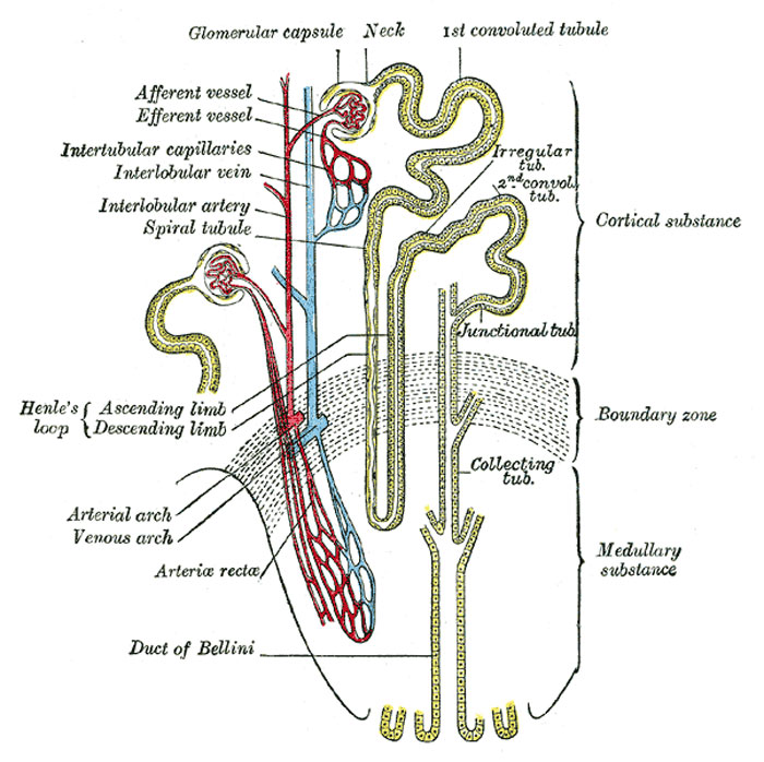 | 700 × 698 (114 KB) | Z8600021 (talk | contribs) | |
| 15:07, 19 September 2009 | 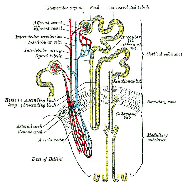 | 602 × 600 (103 KB) | S8600021 (talk | contribs) | Category:Renal Category:Cartoon |
You cannot overwrite this file.
File usage
The following 12 pages use this file:
- 2009 Lecture 15
- 2010 Lecture 15
- ANAT2241 Urinary System
- ANAT2341 Lab 11 - Third Trimester
- ANAT2511 Urinary System
- Anatomy of the Human Body by Henry Gray
- BGDA Practical 12 - Third Trimester
- Lecture - Fetal Development
- Lecture - Renal Development
- Maternal-Fetal Medicine Trainees - Renal
- Renal System Development
- Renal System Histology