File:Gray1095.jpg: Difference between revisions
From Embryology
mNo edit summary |
mNo edit summary |
||
| Line 1: | Line 1: | ||
==Human Gall Bladder== | ==Human Gall Bladder== | ||
The {{ | The {{gallbladder}} (gall-bladder, gallbladder) and bile ducts laid open. (Spalteholz.) | ||
{| | {| | ||
| | | | ||
| Line 28: | Line 28: | ||
[[Category:Gray's 1918 Anatomy]] [[Category:Cartoon]] [[Category:Gastrointestinal Tract]] [[Category: | [[Category:Gray's 1918 Anatomy]] [[Category:Cartoon]] [[Category:Gastrointestinal Tract]] [[Category:Gallbladder]] | ||
Latest revision as of 14:10, 23 January 2019
Human Gall Bladder
The gallbladder (gall-bladder, gallbladder) and bile ducts laid open. (Spalteholz.)
|
|
- Gray's Images: Development | Lymphatic | Neural | Vision | Hearing | Somatosensory | Integumentary | Respiratory | Gastrointestinal | Urogenital | Endocrine | Surface Anatomy | iBook | Historic Disclaimer
| Historic Disclaimer - information about historic embryology pages |
|---|
 |
| iBook - Gray's Embryology | |
|---|---|

|
|
Reference
Gray H. Anatomy of the human body. (1918) Philadelphia: Lea & Febiger.
Cite this page: Hill, M.A. (2024, April 18) Embryology Gray1095.jpg. Retrieved from https://embryology.med.unsw.edu.au/embryology/index.php/File:Gray1095.jpg
- © Dr Mark Hill 2024, UNSW Embryology ISBN: 978 0 7334 2609 4 - UNSW CRICOS Provider Code No. 00098G
File history
Click on a date/time to view the file as it appeared at that time.
| Date/Time | Thumbnail | Dimensions | User | Comment | |
|---|---|---|---|---|---|
| current | 02:05, 8 July 2011 | 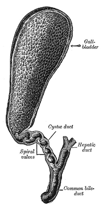 | 349 × 700 (55 KB) | S8600021 (talk | contribs) | ==Human Gall Bladder== * The gall-bladder is a conical or pear-shaped musculomembranous sac * lodged in a fossa on the under surface of the right lobe of the liver * extending from near the right extremity of the porta to the anterior border of the organ |
You cannot overwrite this file.
File usage
The following 3 pages use this file: