File:Gray1015.jpg
Gray1015.jpg (598 × 300 pixels, file size: 62 KB, MIME type: image/jpeg)
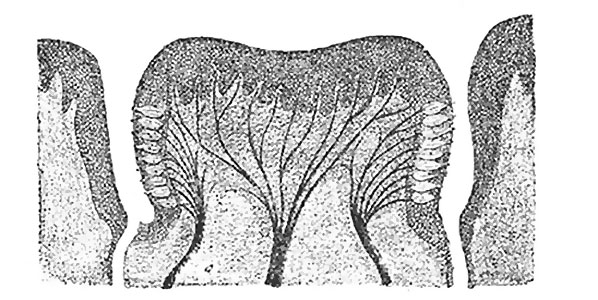
Fig. 1015. Circumvallate papilla in vertical section
Showing arrangement of the taste-buds and nerves.
The papillæ vallatæ (circumvallate papillæ) (Fig. 1015) are of large size, and vary from eight to twelve in number. They are situated on the dorsum of the tongue immediately in front of the foramen cecum and sulcus terminalis, forming a row on either side; the two rows run backward and medialward, and meet in the middle line, like the limbs of the letter V inverted.
Each papilla consists of a projection of mucous membrane from 1 to 2 mm. wide, attached to the bottom of a circular depression of the mucous membrane; the margin of the depression is elevated to form a wall (vallum), and between this and the papilla is a circular sulcus termed the fossa. The papilla is shaped like a truncated cone, the smaller end being directed downward and attached to the tongue, the broader part or base projecting a little above the surface of the tongue and being studded with numerous small secondary papillæ and covered by stratified squamous epithelium.
- Links: Tongue Development | Taste Development | Head Development
- Gray's Images: Development | Lymphatic | Neural | Vision | Hearing | Somatosensory | Integumentary | Respiratory | Gastrointestinal | Urogenital | Endocrine | Surface Anatomy | iBook | Historic Disclaimer
| Historic Disclaimer - information about historic embryology pages |
|---|
| Pages where the terms "Historic" (textbooks, papers, people, recommendations) appear on this site, and sections within pages where this disclaimer appears, indicate that the content and scientific understanding are specific to the time of publication. This means that while some scientific descriptions are still accurate, the terminology and interpretation of the developmental mechanisms reflect the understanding at the time of original publication and those of the preceding periods, these terms, interpretations and recommendations may not reflect our current scientific understanding. (More? Embryology History | Historic Embryology Papers) |
| iBook - Gray's Embryology | |
|---|---|

|
|
Reference
Gray H. Anatomy of the human body. (1918) Philadelphia: Lea & Febiger.
Cite this page: Hill, M.A. (2024, April 25) Embryology Gray1015.jpg. Retrieved from https://embryology.med.unsw.edu.au/embryology/index.php/File:Gray1015.jpg
- © Dr Mark Hill 2024, UNSW Embryology ISBN: 978 0 7334 2609 4 - UNSW CRICOS Provider Code No. 00098G
File history
Click on a date/time to view the file as it appeared at that time.
| Date/Time | Thumbnail | Dimensions | User | Comment | |
|---|---|---|---|---|---|
| current | 08:11, 11 May 2014 |  | 598 × 300 (62 KB) | Z8600021 (talk | contribs) | :'''Links:''' Tongue Development | Taste Development | Head Development | {{Gray Anatomy}} Category:Tongue Category:Head Category:Cartoon |
You cannot overwrite this file.
File usage
The following 2 pages use this file:

