File:Gray0996.jpg: Difference between revisions
(==Adult Palate and Upper Teeth== Permanent teeth of upper dental arch, seen from below. {{Gray Anatomy}} Category:Historic Embryology Category:Gray's 1918 Anatomy Category:Gastrointestinal Tract Category:Palate Category:Tooth) |
|||
| (2 intermediate revisions by the same user not shown) | |||
| Line 1: | Line 1: | ||
==Adult Palate and Upper Teeth== | ==Adult Palate and Upper Teeth== | ||
The Palate (palatum) forms the roof of the mouth; it consists of two portions, the hard palate in front, the soft palate behind. | |||
The Hard Palate (palatum durum) ([[:File:Gray1014.jpg|Fig. 1014]]) is bounded in front and at the sides by the alveolar arches and gums; behind, it is continuous with the soft palate. It is covered by a dense structure, formed by the periosteum and mucous membrane of the mouth, which are intimately adherent. Along the middle line is a linear raphæ, which ends anteriorly in a small papilla corresponding with the incisive canal. On either side and in front of the raphé the mucous membrane is thick, pale in color, and corrugated; behind, it is thin, smooth, and of a deeper color; it is covered with stratified squamous epithelium, and furnished with numerous palatal glands, which lie between the mucous membrane and the surface of the bone. | |||
The Soft Palate (palatum molle) ([[:File:Gray1014.jpg|Fig. 1014]]) is a movable fold, suspended from the posterior border of the hard palate, and forming an incomplete septum between the mouth and pharynx. It consists of a fold of mucous membrane enclosing muscular fibers, an aponeurosis, vessels, nerves, adenoid tissue, and mucous glands. When occupying its usual position, i. e., relaxed and pendent, its anterior surface is concave, continuous with the roof of the mouth, and marked by a median raphé. Its posterior surface is convex, and continuous with the mucous membrane covering the floor of the nasal cavities. Its upper border is attached to the posterior margin of the hard palate, and its sides are blended with the pharynx. Its lower border is free. Its lower portion, which hangs like a curtain between the mouth and pharynx is termed the palatine velum. | |||
Hanging from the middle of its lower border is a small, conical, pendulous process, the palatine uvula; and arching lateralward and downward from the base of the uvula on either side are two curved folds of mucous membrane, containing muscular fibers, called the arches or pillars of the fauces. | |||
{{Palate Links}} | |||
Latest revision as of 09:40, 16 May 2017
Adult Palate and Upper Teeth
The Palate (palatum) forms the roof of the mouth; it consists of two portions, the hard palate in front, the soft palate behind.
The Hard Palate (palatum durum) (Fig. 1014) is bounded in front and at the sides by the alveolar arches and gums; behind, it is continuous with the soft palate. It is covered by a dense structure, formed by the periosteum and mucous membrane of the mouth, which are intimately adherent. Along the middle line is a linear raphæ, which ends anteriorly in a small papilla corresponding with the incisive canal. On either side and in front of the raphé the mucous membrane is thick, pale in color, and corrugated; behind, it is thin, smooth, and of a deeper color; it is covered with stratified squamous epithelium, and furnished with numerous palatal glands, which lie between the mucous membrane and the surface of the bone.
The Soft Palate (palatum molle) (Fig. 1014) is a movable fold, suspended from the posterior border of the hard palate, and forming an incomplete septum between the mouth and pharynx. It consists of a fold of mucous membrane enclosing muscular fibers, an aponeurosis, vessels, nerves, adenoid tissue, and mucous glands. When occupying its usual position, i. e., relaxed and pendent, its anterior surface is concave, continuous with the roof of the mouth, and marked by a median raphé. Its posterior surface is convex, and continuous with the mucous membrane covering the floor of the nasal cavities. Its upper border is attached to the posterior margin of the hard palate, and its sides are blended with the pharynx. Its lower border is free. Its lower portion, which hangs like a curtain between the mouth and pharynx is termed the palatine velum.
Hanging from the middle of its lower border is a small, conical, pendulous process, the palatine uvula; and arching lateralward and downward from the base of the uvula on either side are two curved folds of mucous membrane, containing muscular fibers, called the arches or pillars of the fauces.
| Palate Links: palate | cleft lip and palate | cleft palate | head | Category:Palate |
- Gray's Images: Development | Lymphatic | Neural | Vision | Hearing | Somatosensory | Integumentary | Respiratory | Gastrointestinal | Urogenital | Endocrine | Surface Anatomy | iBook | Historic Disclaimer
| Historic Disclaimer - information about historic embryology pages |
|---|
 |
| iBook - Gray's Embryology | |
|---|---|

|
|
Reference
Gray H. Anatomy of the human body. (1918) Philadelphia: Lea & Febiger.
Cite this page: Hill, M.A. (2024, April 16) Embryology Gray0996.jpg. Retrieved from https://embryology.med.unsw.edu.au/embryology/index.php/File:Gray0996.jpg
- © Dr Mark Hill 2024, UNSW Embryology ISBN: 978 0 7334 2609 4 - UNSW CRICOS Provider Code No. 00098G
File history
Click on a date/time to view the file as it appeared at that time.
| Date/Time | Thumbnail | Dimensions | User | Comment | |
|---|---|---|---|---|---|
| current | 16:16, 28 April 2011 | 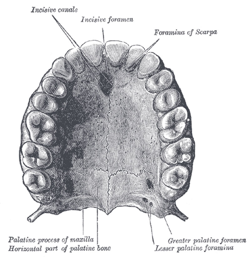 | 800 × 822 (118 KB) | S8600021 (talk | contribs) | ==Adult Palate and Upper Teeth== Permanent teeth of upper dental arch, seen from below. {{Gray Anatomy}} Category:Historic Embryology Category:Gray's 1918 Anatomy Category:Gastrointestinal Tract Category:Palate Category:Tooth |
You cannot overwrite this file.
File usage
The following 2 pages use this file: