File:Gray0987b.jpg
Gray0987b.jpg (554 × 579 pixels, file size: 67 KB, MIME type: image/jpeg)
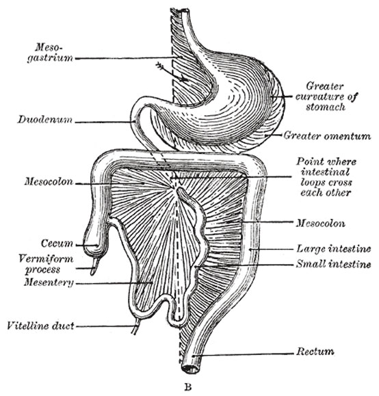
Development of the Digestive Tube and its Mesentery
Diagrams to illustrate two stages in the development of the digestive tube and its mesentery. The arrow indicates the entrance to the bursa omentalis.
At this stage the small and large intestines are attached to the vertebral column by a common mesentery, the coils of the small intestine falling to the right of the middle line, while the large intestine lies on the left side.
The gut is now rotated upon itself, so that the large intestine is carried over in front of the small intestine, and the cecum is placed immediately below the liver; about the sixth month the cecum descends into the right iliac fossa, and the large intestine forms an arch consisting of the ascending, transverse, and descending portions of the colon—the transverse portion crossing in front of the duodenum and lying just below the greater curvature of the stomach; within this arch the coils of the small intestine are disposed (Fig. 988). Sometimes the downward progress of the cecum is arrested, so that in the adult it may be found lying immediately below the liver instead of in the right iliac region.
Further changes take place in the bursa omentalis and in the common mesentery, and give rise to the peritoneal relations seen in the adult. The bursa omentalis, which at first reaches only as far as the greater curvature of the stomach, grows downward to form the greater omentum, and this downward extension lies in front of the transverse colon and the coils of the small intestine (Fig. 989). Above, before the pleuro-peritoneal opening is closed, the bursa omentalis sends up a diverticulum on either side of the esophagus; the left diverticulum soon disappears, but the right is constricted off and persists in most adults as a small sac lying within the thorax on the right side of the lower end of the esophagus. The anterior layer of the transverse mesocolon is at first distinct from the posterior layer of the greater omentum, but ultimately the two blend, and hence the greater omentum appears as if attached to the transverse colon (Fig. 990). The mesenteries of the ascending and descending parts of the colon disappear in the majority of cases, while that of the small intestine assumes the oblique attachment characteristic of its adult condition.
- Links: Image 987a | Image 987b | Image - Early Week 4 | Image - Late Week 4 | Gastrointestinal Tract Development | Endoderm
- Gray's Images: Development | Lymphatic | Neural | Vision | Hearing | Somatosensory | Integumentary | Respiratory | Gastrointestinal | Urogenital | Endocrine | Surface Anatomy | iBook | Historic Disclaimer
| Historic Disclaimer - information about historic embryology pages |
|---|
 |
| iBook - Gray's Embryology | |
|---|---|

|
|
Reference
Gray H. Anatomy of the human body. (1918) Philadelphia: Lea & Febiger.
Cite this page: Hill, M.A. (2024, April 19) Embryology Gray0987b.jpg. Retrieved from https://embryology.med.unsw.edu.au/embryology/index.php/File:Gray0987b.jpg
- © Dr Mark Hill 2024, UNSW Embryology ISBN: 978 0 7334 2609 4 - UNSW CRICOS Provider Code No. 00098G
File history
Click on a date/time to view the file as it appeared at that time.
| Date/Time | Thumbnail | Dimensions | User | Comment | |
|---|---|---|---|---|---|
| current | 15:02, 28 April 2011 | | 554 × 579 (67 KB) | S8600021 (talk | contribs) | ==Development of the Digestive Tube and its Mesentery== Diagrams to illustrate two stages in the development of the digestive tube and its mesentery. The arrow indicates the entrance to the bursa omentalis. At this stage the small and large intestines a |
You cannot overwrite this file.
File usage
The following 2 pages use this file: