File:Gray0982b.jpg
From Embryology
No higher resolution available.
Gray0982b.jpg (427 × 393 pixels, file size: 20 KB, MIME type: image/jpeg)
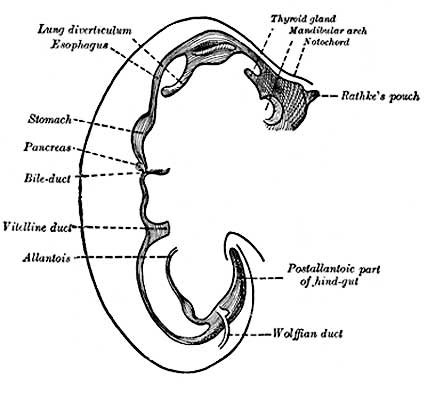
Sketches in profile of two stages in the development of the human digestive tube. (His.) A X 30. B X 20.
- Gray's Images: Development | Lymphatic | Neural | Vision | Hearing | Somatosensory | Integumentary | Respiratory | Gastrointestinal | Urogenital | Endocrine | Surface Anatomy | iBook | Historic Disclaimer
| Historic Disclaimer - information about historic embryology pages |
|---|
 |
| iBook - Gray's Embryology | |
|---|---|

|
|
Reference
Gray H. Anatomy of the human body. (1918) Philadelphia: Lea & Febiger.
Cite this page: Hill, M.A. (2024, April 24) Embryology Gray0982b.jpg. Retrieved from https://embryology.med.unsw.edu.au/embryology/index.php/File:Gray0982b.jpg
- © Dr Mark Hill 2024, UNSW Embryology ISBN: 978 0 7334 2609 4 - UNSW CRICOS Provider Code No. 00098G
Image Source: Anatomy of the Human Body (1918) by Henry Gray
File history
Click on a date/time to view the file as it appeared at that time.
| Date/Time | Thumbnail | Dimensions | User | Comment | |
|---|---|---|---|---|---|
| current | 15:34, 23 August 2009 | | 427 × 393 (20 KB) | S8600021 (talk | contribs) |
You cannot overwrite this file.
File usage
The following 20 pages use this file:
- 2009 Lecture 10
- 2009 Lecture 9
- 2010 Lecture 10
- 2010 Lecture 9
- 2011 Lab 5 - Early Embryo
- ANAT2341 Lab 5 - Early Embryo
- Anatomy of the Human Body by Henry Gray
- BGDB Gastrointestinal - Activity 2
- BGDB Gastrointestinal - Early Embryo
- Draft 2016
- Gastrointestinal Tract - Mouth Development
- Gastrointestinal Tract - Oesophagus Development
- Gastrointestinal Tract Development
- Lecture - Gastrointestinal Development
- Lecture - Gastrointestinal Development 2013
- Lecture - Respiratory Development
- Respiratory System - Upper Respiratory Tract
- Respiratory System Development
- SH Lecture - Respiratory System Development
- Talk:2010 Lecture 9