File:Gray0974.jpg
From Embryology
Size of this preview: 462 × 599 pixels. Other resolution: 617 × 800 pixels.
Original file (617 × 800 pixels, file size: 125 KB, MIME type: image/jpeg)
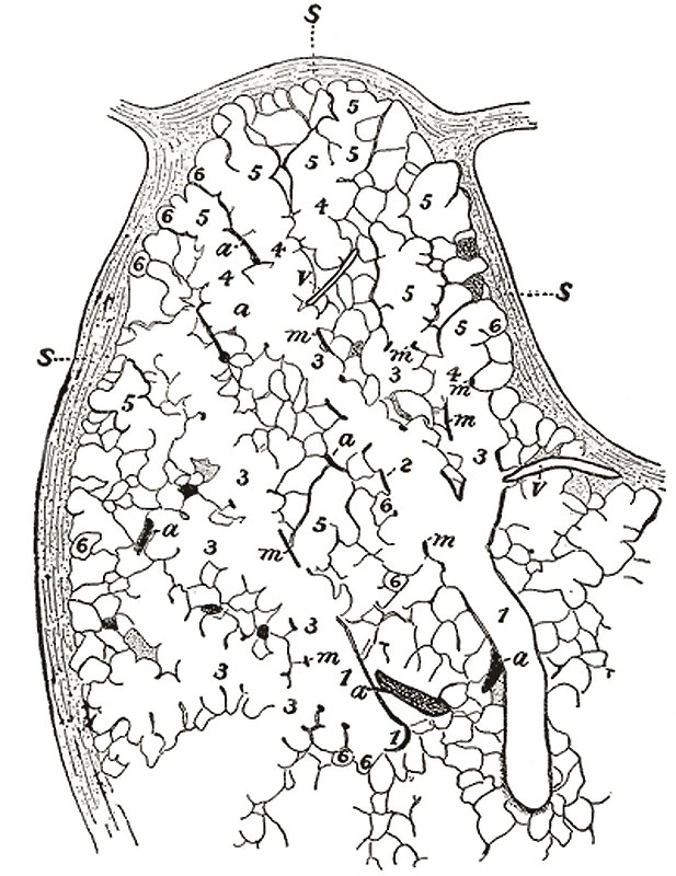
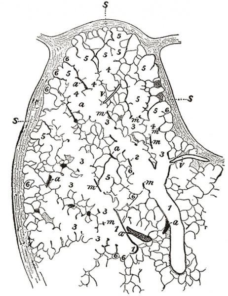
Lung Anatomy - Secondary Lobule
Part of a secondary lobule from the depth of a human lung, showing parts of several primary lobules.
Camera drawing of one 50 μm section. X 20 diameters. (Miller.)
|
|
See also Labeled version
- Gray's Images: Development | Lymphatic | Neural | Vision | Hearing | Somatosensory | Integumentary | Respiratory | Gastrointestinal | Urogenital | Endocrine | Surface Anatomy | iBook | Historic Disclaimer
| Historic Disclaimer - information about historic embryology pages |
|---|
 |
| iBook - Gray's Embryology | |
|---|---|

|
|
Reference
Gray H. Anatomy of the human body. (1918) Philadelphia: Lea & Febiger.
Cite this page: Hill, M.A. (2024, April 24) Embryology Gray0974.jpg. Retrieved from https://embryology.med.unsw.edu.au/embryology/index.php/File:Gray0974.jpg
- © Dr Mark Hill 2024, UNSW Embryology ISBN: 978 0 7334 2609 4 - UNSW CRICOS Provider Code No. 00098G
File history
Click on a date/time to view the file as it appeared at that time.
| Date/Time | Thumbnail | Dimensions | User | Comment | |
|---|---|---|---|---|---|
| current | 16:36, 29 February 2012 | | 617 × 800 (125 KB) | Z8600021 (talk | contribs) | |
| 20:40, 24 August 2009 |  | 462 × 600 (58 KB) | S8600021 (talk | contribs) | Part of a secondary lobule from the depth of a human lung, showing parts of several primary lobules. Camera drawing of one 50 μ section. X 20 diameters. (Miller.) # bronchiole # respiratory bronchiole # alveolar duct # atria # alveolar sac # alveolus o |
You cannot overwrite this file.