File:Gray0974.jpg: Difference between revisions
From Embryology
No edit summary |
|||
| Line 4: | Line 4: | ||
Camera drawing of one 50 μm section. X 20 diameters. (Miller.) | Camera drawing of one 50 μm section. X 20 diameters. (Miller.) | ||
{| | |||
| | |||
# bronchiole | # bronchiole | ||
# respiratory bronchiole | # respiratory bronchiole | ||
| Line 11: | Line 12: | ||
# alveolar sac | # alveolar sac | ||
# alveolus or air cell | # alveolus or air cell | ||
| | |||
* '''m''' - smooth muscle | * '''m''' - smooth muscle | ||
* '''a''' - branch pulmonary artery | * '''a''' - branch pulmonary artery | ||
* '''v''' - branch pulmonary vein | * '''v''' - branch pulmonary vein | ||
* '''s''' - septum between secondary lobules | * '''s''' - septum between secondary lobules | ||
|} | |||
{{Grays Anatomy}} | |||
[[Category:Respiratory]] [[Category:Historic Embryology]] [[Category:Gray's 1918 Anatomy]] | [[Category:Respiratory]] [[Category:Historic Embryology]] [[Category:Gray's 1918 Anatomy]] | ||
Revision as of 03:00, 17 August 2012
Lung Anatomy - Secondary Lobule
Part of a secondary lobule from the depth of a human lung, showing parts of several primary lobules.
Camera drawing of one 50 μm section. X 20 diameters. (Miller.)
|
|
File history
Click on a date/time to view the file as it appeared at that time.
| Date/Time | Thumbnail | Dimensions | User | Comment | |
|---|---|---|---|---|---|
| current | 16:36, 29 February 2012 | 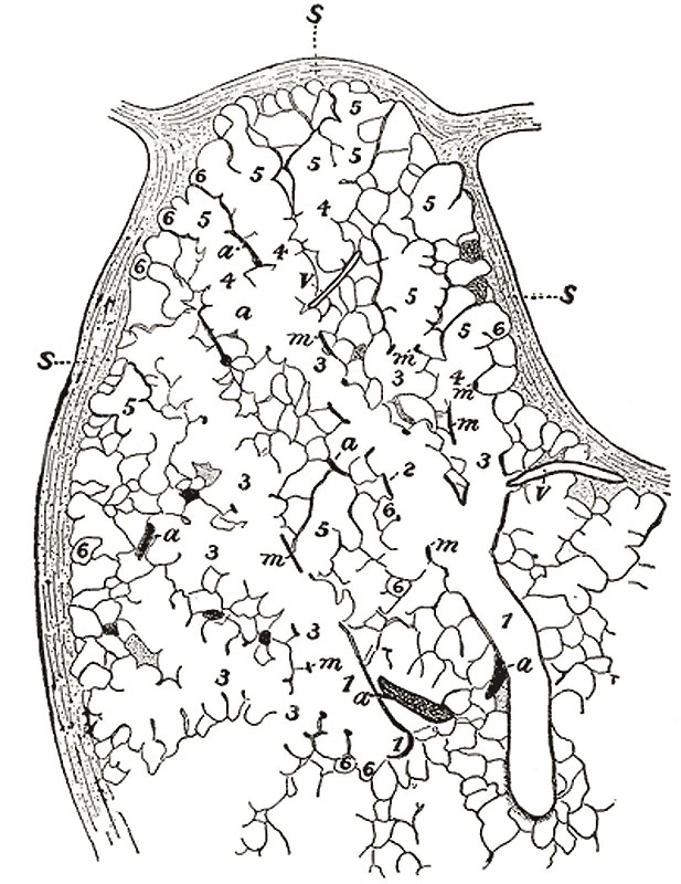 | 617 × 800 (125 KB) | Z8600021 (talk | contribs) | |
| 20:40, 24 August 2009 |  | 462 × 600 (58 KB) | S8600021 (talk | contribs) | Part of a secondary lobule from the depth of a human lung, showing parts of several primary lobules. Camera drawing of one 50 μ section. X 20 diameters. (Miller.) # bronchiole # respiratory bronchiole # alveolar duct # atria # alveolar sac # alveolus o |
You cannot overwrite this file.