File:Gray0924.jpg
Gray0924.jpg (500 × 384 pixels, file size: 26 KB, MIME type: image/jpeg)
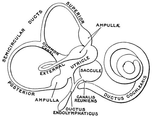
Fig. 924. Inner Ear - The Membranous Labyrinth
The Membranous Labyrinth
(labyrinthus membranaceus) (Figs. 924, 925, 926) — The membranous labyrinth is lodged within the bony cavities just described, and has the same general form as these; it is, however, considerably smaller, and is partly separated from the bony walls by a quantity of fluid, the perilymph. In certain places it is fixed to the walls of the cavity. The membranous labyrinth contains fluid, the endolymph, and on its walls the ramifications of the acoustic nerve are distributed.
Within the osseous vestibule the membranous labyrinth does not quite preserve the form of the bony cavity, but consists of two membranous sacs, the utricle, and the saccule.
The Utricle
(utriculus) The utricle, the larger of the two, is of an oblong form, compressed transversely, and occupies the upper and back part of the vestibule, lying in contact with the recessus ellipticus and the part below it. That portion which is lodged in the recess forms a sort of pouch or cul-de-sac, the floor and anterior wall of which are thickened, and form the macula acustica utriculi, which receives the utricular filaments of the acoustic nerve. The cavity of the utricle communicates behind with the semicircular ducts by five orifices. From its anterior wall is given off the ductus utriculosaccularis, which opens into the ductus endolymphaticus.
The Saccule
(sacculus) The saccule is the smaller of the two vestibular sacs; it is globular in form, and lies in the recessus sphæricus near the opening of the scala vestibuli of the cochlea. Its anterior part exhibits an oval thickening, the macula acustica sacculi, to which are distributed the saccular filaments of the acoustic nerve. Its cavity does not directly communicate with that of the utricle. From the posterior wall a canal, the ductus endolymphaticus, is given off; this duct is joined by the ductus utriculosaccularis, and then passes along the aquæductus vestibuli and ends in a blind pouch (saccus endolymphaticus) on the posterior surface of the petrous portion of the temporal bone, where it is in contact with the dura mater. From the lower part of the saccule a short tube, the canalis reuniens of Hensen, passes downward and opens into the ductus cochlearis near its vestibular extremity (Fig. 924).
The Semicircular Ducts
(ductus semicirculares; membranous semicircular canals), (Figs. 925, 926).—The semicircular ducts are about one-fourth of the diameter of the osseous canals, but in number, shape, and general form they are precisely similar, and each presents at one end an ampulla. They open by five orifices into the utricle, one opening being common to the medial end of the superior and the upper end of the posterior duct. In the ampullæ the wall is thickened, and projects into the cavity as a fiddle-shaped, transversely placed elevation, the septum transversum, in which the nerves end.
The utricle, saccule, and semicircular ducts are held in position by numerous fibrous bands which stretch across the space between them and the bony walls.
(text modified from Gray's 1918 Anatomy)
- Links: Balance Development | Inner Ear Development | Hearing and Balance Development | Gray's 1918 Anatomy
- Gray's Images: Development | Lymphatic | Neural | Vision | Hearing | Somatosensory | Integumentary | Respiratory | Gastrointestinal | Urogenital | Endocrine | Surface Anatomy | iBook | Historic Disclaimer
| Historic Disclaimer - information about historic embryology pages |
|---|
 |
| iBook - Gray's Embryology | |
|---|---|

|
|
Reference
Gray H. Anatomy of the human body. (1918) Philadelphia: Lea & Febiger.
Cite this page: Hill, M.A. (2024, April 19) Embryology Gray0924.jpg. Retrieved from https://embryology.med.unsw.edu.au/embryology/index.php/File:Gray0924.jpg
- © Dr Mark Hill 2024, UNSW Embryology ISBN: 978 0 7334 2609 4 - UNSW CRICOS Provider Code No. 00098G
File history
Click on a date/time to view the file as it appeared at that time.
| Date/Time | Thumbnail | Dimensions | User | Comment | |
|---|---|---|---|---|---|
| current | 00:16, 28 September 2009 | | 500 × 384 (26 KB) | S8600021 (talk | contribs) |
You cannot overwrite this file.