File:Gray0916.jpg
From Embryology
No higher resolution available.
Gray0916.jpg (600 × 367 pixels, file size: 28 KB, MIME type: image/jpeg)
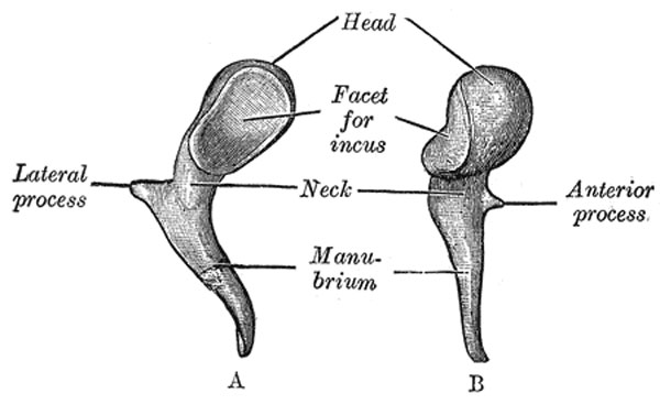
Fig. 916 Middle Ear - Malleus
Left malleus. A. From behind. B. From within.
- named from its fancied resemblance to a hammer
- consists of a head, neck, and three processes
- the manubrium, the anterior and lateral processes
Hearing - Middle Ear Development
Head (capitulum mallei)
- large upper extremity of the bone
- oval in shape
- articulates posteriorly with the incus
- being free in the rest of its extent
- facet for articulation with the incus is constricted near the middle
- consists of an upper larger and lower smaller part
- form nearly a right angle with each other.
- opposite the constriction
- lower margin of the facet projects in the form of a process (cog-tooth or spur of the malleus)
Neck (collum mallei)
- narrow contracted part
- just beneath the head
- below it is a prominence
- which the various processes are attached
Handle (manubrium mallei)
- connected by its lateral margin with the tympanic membrane
- directed downward, medialward, and backward
- decreases in size toward its free end, which is curved slightly forward, and flattened transversely
- medial side, near its upper end, is a slight projection, into which the tendon of the tensor tympani is inserted
Anterior Process (processus anterior [Folii]; processus gracilis)
- delicate spicule from the eminence below the neck
- directed forward to the petrotympanic fissure
- to which it is connected by ligamentous fibers
In the fetus this is the longest process of the malleus, and is in direct continuity with Meckel's cartilage.
Lateral Process (processus lateralis; processus brevis)
- a slight conical projection from the root of the manubrium
- directed laterally, and is attached to the upper part of the tympanic membrane
- by means of the anterior and posterior malleolar folds, to the extremities of the notch of Rivinus.
(text modified from Gray's Anatomy)
- Gray's Images: Development | Lymphatic | Neural | Vision | Hearing | Somatosensory | Integumentary | Respiratory | Gastrointestinal | Urogenital | Endocrine | Surface Anatomy | iBook | Historic Disclaimer
| Historic Disclaimer - information about historic embryology pages |
|---|
 |
| iBook - Gray's Embryology | |
|---|---|

|
|
Reference
Gray H. Anatomy of the human body. (1918) Philadelphia: Lea & Febiger.
Cite this page: Hill, M.A. (2024, April 23) Embryology Gray0916.jpg. Retrieved from https://embryology.med.unsw.edu.au/embryology/index.php/File:Gray0916.jpg
- © Dr Mark Hill 2024, UNSW Embryology ISBN: 978 0 7334 2609 4 - UNSW CRICOS Provider Code No. 00098G
Development
Human malleus development timing:[1]
- 16 weeks - two cortical fascicles situated in the neck
- 21 weeks - fascicles extend towards the head
- 23 weeks - extend towards to the lateral process
- 24 weeks - extend towards to the handle
- 29 weeks - in the handle force lines are transmitted via three cardinal fascicles (two of them via the cortical fascicle and one via the centre)
- 31 weeks - consolidated by this time
- ↑ <pubmed>18581276</pubmed>
File history
Click on a date/time to view the file as it appeared at that time.
| Date/Time | Thumbnail | Dimensions | User | Comment | |
|---|---|---|---|---|---|
| current | 02:33, 20 May 2011 | | 600 × 367 (28 KB) | S8600021 (talk | contribs) | ==Middle Ear - Malleus== Left malleus. A. From behind. B. From within. {{Gray Anatomy}} Category:Hearing |
You cannot overwrite this file.
File usage
The following 5 pages use this file: