File:Gray0848.jpg
Original file (800 × 935 pixels, file size: 289 KB, MIME type: image/jpeg)
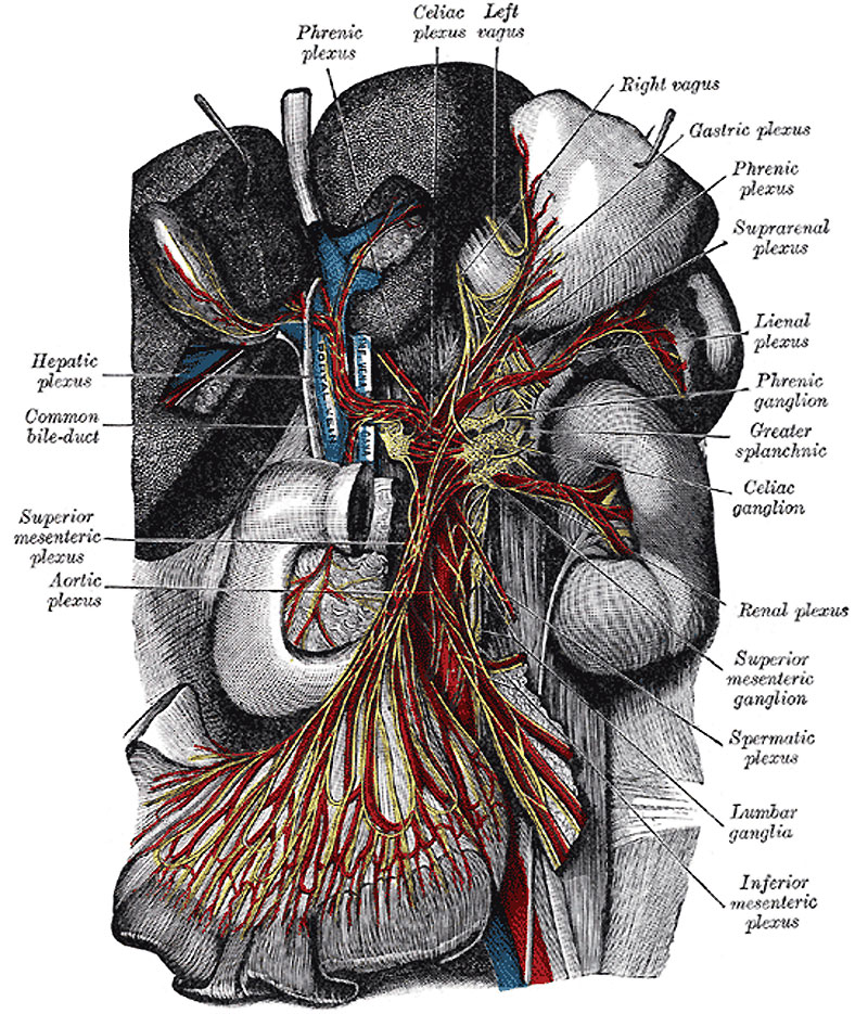
The Great Plexuses of the Sympathetic System
The celiac ganglia with the sympathetic plexuses of the abdominal viscera radiating from the ganglia. (Toldt.)
- Links: Neural Crest | Cardiovascular | Gastrointestinal Tract | Renal
Text below modified from Gray's Anatomy, 1918.
The great plexuses of the sympathetic are aggregations of nerves and ganglia, situated in the thoracic, abdominal, and pelvic cavities, and named the cardiac, celiac, and hypogastric plexuses. They consist not only of sympathetic fibers derived from the ganglia, but of fibers from the medulla spinalis, which are conveyed through the white rami communicantes. From the plexuses branches are given to the thoracic, abdominal, and pelvic viscera.
The Cardiac Plexus
The Cardiac Plexus (Plexus Cardiacus) - The cardiac plexus is situated at the base of the heart, and is divided into a superficial part, which lies in the concavity of the aortic arch, and a deep part, between the aortic arch and the trachea. The two parts are, however, closely connected.
The superficial part of the cardiac plexus lies beneath the arch of the aorta, in front of the right pulmonary artery. It is formed by the superior cardiac branch of the left sympathetic and the lower superior cervical cardiac branch of the left vagus. A small ganglion, the cardiac ganglion of Wrisberg, is occasionally found connected with these nerves at their point of junction. This ganglion, when present, is situated immediately beneath the arch of the aorta, on the right side of the ligamentum arteriosum. The superficial part of the cardiac plexus gives branches (a) to the deep part of the plexus; (b) to the anterior coronary plexus; and (c) to the left anterior pulmonary plexus.
The deep part of the cardiac plexus is situated in front of the bifurcation of the trachea, above the point of division of the pulmonary artery, and behind the aortic arch. It is formed by the cardiac nerves derived from the cervical ganglia of the sympathetic, and the cardiac branches of the vagus and recurrent nerves. The only cardiac nerves which do not enter into the formation of the deep part of the cardiac plexus are the superior cardiac nerve of the left sympathetic, and the lower of the two superior cervical cardiac branches from the left vagus, which pass to the superficial part of the plexus.
The branches from the right half of the deep part of the cardiac plexus pass, some in front of, and others behind, the right pulmonary artery; the former, the more numerous, transmit a few filaments to the anterior pulmonary plexus, and are then continued onward to form part of the anterior coronary plexus; those behind the pulmonary artery distribute a few filaments to the right atrium, and are then continued onward to form part of the posterior coronary plexus.
The left half of the deep part of the plexus is connected with the superficial part of the cardiac plexus, and gives filaments to the left atrium, and to the anterior pulmonary plexus, and is then continued to form the greater part of the posterior coronary plexus.
Posterior Coronary Plexus
The Posterior Coronary Plexus (plexus coronarius posterior; left coronary plexus) is larger than the anterior, and accompanies the left coronary artery; it is chiefly formed by filaments prolonged from the left half of the deep part of the cardiac plexus, and by a few from the right half. It gives branches to the left atrium and ventricle.
Anterior Coronary Plexus
The Anterior Coronary Plexus (plexus coronarius anterior; right coronary plexus) is formed partly from the superficial and partly from the deep parts of the cardiac plexus. It accompanies the right coronary artery, and gives branches to the right atrium and ventricle.
The Celiac Plexus
The Celiac Plexus (Plexus Cœliacus; Solar Plexus) (Figs. 838, 848) - The celiac plexus, the largest of the three sympathetic plexuses, is situated at the level of the upper part of the first lumbar vertebra and is composed of two large ganglia, the celiac ganglia, and a dense net-work of nerve fibers uniting them together. It surrounds the celiac artery and the root of the superior mesenteric artery. It lies behind the stomach and the omental bursa, in front of the crura of the diaphragm and the commencement of the abdominal aorta, and between the suprarenal glands. The plexus and the ganglia receive the greater and lesser splanchnic nerves of both sides and some filaments from the right vagus, and give off numerous secondary plexuses along the neighboring arteries.
Celiac Ganglia
The Celiac Ganglia (ganglia cæliaca; semilunar ganglia) are two large irregularly shaped masses having the appearance of lymph glands and placed one on either side of the middle line in front of the crura of the diaphragm close to the suprarenal glands, that on the right side being placed behind the inferior vena cava. The upper part of each ganglion is joined by the greater splanchnic nerve, while the lower part, which is segmented off and named the aorticorenal ganglion, receives the lesser splanchnic nerve and gives off the greater part of the renal plexus.
The secondary plexuses springing from or connected with the celiac plexus are the
- Phrenic.
- Renal.
- Hepatic.
- Spermatic.
- Lienal.
- Superior mesenteric.
- Superior gastric.
- Abdominal aortic.
- Suprarenal.
- Inferior mesenteric.
Phrenic Plexus
The phrenic plexus (plexus phrenicus) accompanies the inferior phrenic artery to the diaphragm, some filaments passing to the suprarenal gland. It arises from the upper part of the celiac ganglion, and is larger on the right than on the left side. It receives one or two branches from the phrenic nerve. At the point of junction of the right phrenic plexus with the phrenic nerve is a small ganglion (ganglion phrenicum). This plexus distributes branches to the inferior vena cava, and to the suprarenal and hepatic plexuses.
Hepatic Plexus
The hepatic plexus (plexus hepaticus), the largest offset from the celiac plexus, receives filaments from the left vagus and right phrenic nerves. It accompanies the hepatic artery, ramifying upon its branches, and upon those of the portal vein in the substance of the liver. Branches from this plexus accompany all the divisions of the hepatic artery. A considerable plexus accompanies the gastroduodenal artery and is continued as the inferior gastric plexus on the right gastroepiploic artery along the greater curvature of the stomach, where it unites with offshoots from the lienal plexus.
Lienal Plexus
The lienal plexus (plexus lienalis; splenic plexus) is formed by branches from the celiac plexus, the left celiac ganglion, and from the right vagus nerve. It accompanies the lienal artery to the spleen, giving off, in its course, subsidiary plexuses along the various branches of the artery.
Superior Gastric Plexus
The superior gastric plexus (plexus gastricus superior; gastric or coronary plexus) accompanies the left gastric artery along the lesser curvature of the stomach, and joins with branches from the left vagus.
Suprarenal Plexus
The suprarenal plexus (plexus suprarenalis) is formed by branches from the celiac plexus, from the celiac ganglion, and from the phrenic and greater splanchnic nerves, a ganglion being formed at the point of junction with the latter nerve. The plexus supplies the suprarenal gland, being distributed chiefly to its medullary portion; its branches are remarkable for their large size in comparison with that of the organ they supply.
Renal Plexus
The renal plexus (plexus renalis) is formed by filaments from the celiac plexus, the aorticorenal ganglion, and the aortic plexus. It is joined also by the smallest splanchnic nerve. The nerves from these sources, fifteen or twenty in number, have a few ganglia developed upon them. They accompany the branches of the renal artery into the kidney; some filaments are distributed to the spermatic plexus and, on the right side, to the inferior vena cava.
Spermatic Plexus
The spermatic plexus (plexus spermaticus) is derived from the renal plexus, receiving branches from the aortic plexus. It accompanies the internal spermatic artery to the testis. In the female, the ovarian plexus (plexus arteriæ ovaricæ) arises from the renal plexus, and is distributed to the ovary, and fundus of the uterus.
Superior Mesenteric Plexus
The superior mesenteric plexus (plexus mesentericus superior) is a continuation of the lower part of the celiac plexus, receiving a branch from the junction of the right vagus nerve with the plexus. It surrounds the superior mesenteric artery, accompanies it into the mesentery, and divides into a number of secondary plexuses, which are distributed to all the parts supplied by the artery, viz., pancreatic branches to the pancreas; intestinal branches to the small intestine; and ileocolic, right colic, and middle colic branches, which supply the corresponding parts of the great intestine. The nerves composing this plexus are white in color and firm in texture; in the upper part of the plexus close to the origin of the superior mesenteric artery is a ganglion (ganglion mesentericum superius).
Abdominal Aortic Plexus
The abdominal aortic plexus (plexus aorticus abdominalis; aortic plexus) is formed by branches derived, on either side, from the celiac plexus and ganglia, and receives filaments from some of the lumbar ganglia. It is situated upon the sides and front of the aorta, between the origins of the superior and inferior mesenteric arteries. From this plexus arise part of the spermatic, the inferior mesenteric, and the hypogastric plexuses; it also distributes filaments to the inferior vena cava.
The inferior mesenteric plexus (plexus mesentericus inferior) is derived chiefly from the aortic plexus. It surrounds the inferior mesenteric artery, and divides into a number of secondary plexuses, which are distributed to all the parts supplied by the artery, viz., the left colic and sigmoid plexuses, which supply the descending and sigmoid parts of the colon; and the superior hemorrhoidal plexus, which supplies the rectum and joins in the pelvis with branches from the pelvic plexuses.
Hypogastric Plexus
The Hypogastric Plexus (Plexus Hypogastricus) - The hypogastric plexus is situated in front of the last lumbar vertebra and the promontory of the sacrum, between the two common iliac arteries, and is formed by the union of numerous filaments, which descend on either side from the aortic plexus, and from the lumbar ganglia; it divides, below, into two lateral portions which are named the pelvic plexuses.
Pelvic Plexuses
The pelvic plexuses supply the viscera of the pelvic cavity, and are situated at the sides of the rectum in the male, and at the sides of the rectum and vagina in the female. They are formed on either side by a continuation of the hypogastric plexus, by the sacral sympathetic efferent fibers from the second, third, and fourth sacral nerves, and by a few filaments from the first two sacral ganglia. At the points of junction of these nerves small ganglia are found. From these plexuses numerous branches are distributed to the viscera of the pelvis. They accompany the branches of the hypogastric artery.
Middle Hemorrhoidal Plexus
The Middle Hemorrhoidal Plexus (plexus hæmorrhoidalis medius) arises from the upper part of the pelvic plexus. It supplies the rectum, and joins with branches of the superior hemorrhoidal plexus.
(Text from Gray's Anatomy, 1918)
- Gray's Images: Development | Lymphatic | Neural | Vision | Hearing | Somatosensory | Integumentary | Respiratory | Gastrointestinal | Urogenital | Endocrine | Surface Anatomy | iBook | Historic Disclaimer
| Historic Disclaimer - information about historic embryology pages |
|---|
 |
| iBook - Gray's Embryology | |
|---|---|

|
|
Reference
Gray H. Anatomy of the human body. (1918) Philadelphia: Lea & Febiger.
Cite this page: Hill, M.A. (2024, April 19) Embryology Gray0848.jpg. Retrieved from https://embryology.med.unsw.edu.au/embryology/index.php/File:Gray0848.jpg
- © Dr Mark Hill 2024, UNSW Embryology ISBN: 978 0 7334 2609 4 - UNSW CRICOS Provider Code No. 00098G
File history
Click on a date/time to view the file as it appeared at that time.
| Date/Time | Thumbnail | Dimensions | User | Comment | |
|---|---|---|---|---|---|
| current | 12:11, 29 April 2011 | | 800 × 935 (289 KB) | S8600021 (talk | contribs) | better text resolution. |
| 11:31, 29 April 2011 | 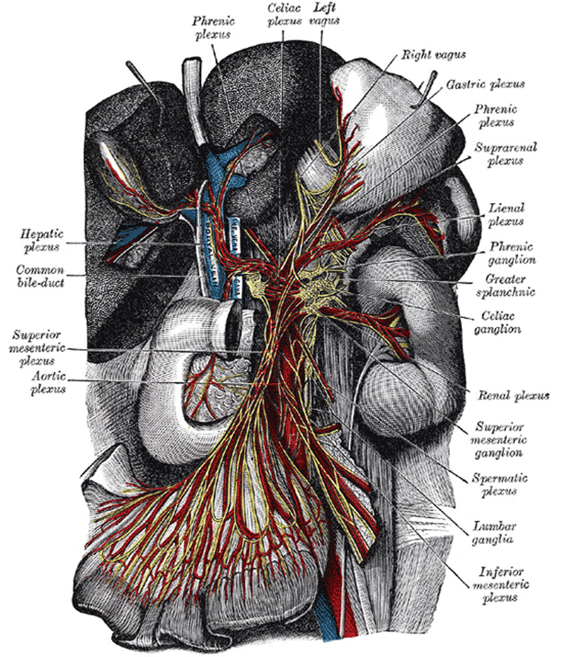 | 800 × 935 (245 KB) | S8600021 (talk | contribs) | ==The Great Plexuses of the Sympathetic System== The celiac ganglia with the sympathetic plexuses of the abdominal viscera radiating from the ganglia. (Toldt.) Text below modified from Gray's Anatomy, 1918. The great plexuses of the sympathetic are ag |
You cannot overwrite this file.