File:Gray0475.jpg
Original file (2,042 × 1,363 pixels, file size: 350 KB, MIME type: image/jpeg)
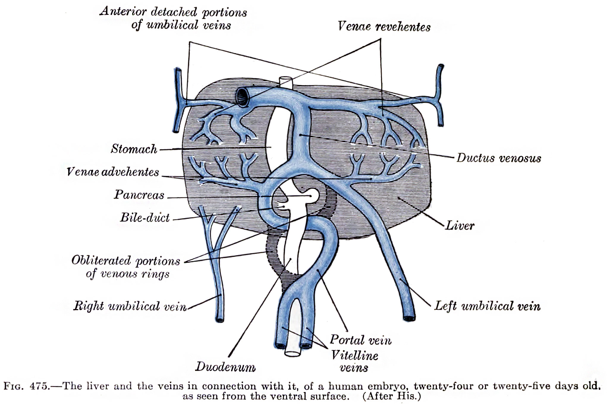
Human Embryo Liver and Associated Veins
Human embryo, 24 or 25 days old (liver ventral surface view) Figure after His.
- ductus venosus - shunts approximately half the umbilical vein blood flow directly to the inferior vena cava.
- portal vein - carries blood from the gastrointestinal tract and spleen to the liver.
- left umbilical vein - carries oxygenated blood from the placenta to the embryo/fetus.
- right umbilical vein - vessel degenerates leaving a single (left) umbilical vein.
- vena revehentes - veins from the sinusoid vessels in the liver to the inferior vena cava, that later develop into the hepatic veins.
- Links: Liver | Cardiovascular System Development
The Vitelline Veins run upward at first in front, and subsequently on either side of the intestinal canal. They unite on the ventral aspect of the canal, and beyond this are connected to one another by two anastomotic branches, one on the dorsal, and the other on the ventral aspect of the duodenal portion of the intestine, which is thus encircled by two venous rings (Fig. 475); into the middle or dorsal anastomosis the superior mesenteric vein opens. The portions of the veins above the upper ring become interrupted by the developing liver and broken up by it into a plexus of small capillary-like vessels termed sinusoids (Minot). The branches conveying the blood to this plexus are named the venæ advehentes, and become the branches of the portal vein; while the vessels draining the plexus into the sinus venosus are termed the venæ revehentes, and form the future hepatic veins (Figs. 475, 476). Ultimately the left vena revehens no longer communicates directly with the sinus venosus, but opens into the right vena revehens. The persistent part of the upper venous ring, above the opening of the superior mesenteric vein, forms the trunk of the portal vein.
(Gray's text)
- Gray - Cardiovascular: 448 Artery and vein | 458 Human embryo week 2 vascular | 459 Human embryo 14 days with yolk-sac
- Gray's Images: Development | Lymphatic | Neural | Vision | Hearing | Somatosensory | Integumentary | Respiratory | Gastrointestinal | Urogenital | Endocrine | Surface Anatomy | iBook | Historic Disclaimer
| Historic Disclaimer - information about historic embryology pages |
|---|
 |
| iBook - Gray's Embryology | |
|---|---|

|
|
Reference
Gray H. Anatomy of the human body. (1918) Philadelphia: Lea & Febiger.
Cite this page: Hill, M.A. (2024, April 19) Embryology Gray0475.jpg. Retrieved from https://embryology.med.unsw.edu.au/embryology/index.php/File:Gray0475.jpg
- © Dr Mark Hill 2024, UNSW Embryology ISBN: 978 0 7334 2609 4 - UNSW CRICOS Provider Code No. 00098G
File history
Click on a date/time to view the file as it appeared at that time.
| Date/Time | Thumbnail | Dimensions | User | Comment | |
|---|---|---|---|---|---|
| current | 16:25, 21 February 2015 | | 2,042 × 1,363 (350 KB) | Z8600021 (talk | contribs) | |
| 22:20, 11 October 2009 |  | 600 × 484 (44 KB) | S8600021 (talk | contribs) | The liver and the veins in connection with it, of a human embryo, twenty-four or twenty-five days old, as seen from the ventral surface. (After His.) |
You cannot overwrite this file.
File usage
The following 3 pages use this file: