File:Gray0185.jpg
Gray0185.jpg (600 × 491 pixels, file size: 36 KB, MIME type: image/jpeg)
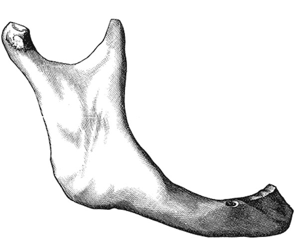
Human Mandible in Old Age
In old age (Fig. 185) the bone becomes greatly reduced in size, for with the loss of the teeth the alveolar process is absorbed, and, consequently, the chief part of the bone is below the oblique line. The mandibular canal, with the mental foramen opening from it, is close to the alveolar border. The ramus is oblique in direction, the angle measures about 140o, and the neck of the condyle is more or less bent backward.
- Mandible Development: Week 8 outer view | Week 8 inner view | Week 12 outer view | Week 12 inner view | Week 12 Head outer view | Week 12 Head inner view | Birth | Childhood | Adult | Old Age | Small Animation | Large Animation | Muscle Attachments | Mandible Ossification | 1909 Mandible | embryo 18 mm | embryo 24 mm | embryo 28 mm | fetus 43 mm | fetus 65 mm | fetus 55 mm | fetus 95 mm | human 18-24-95 mm | Skull Development | Head Development
- Gray's Images: Development | Lymphatic | Neural | Vision | Hearing | Somatosensory | Integumentary | Respiratory | Gastrointestinal | Urogenital | Endocrine | Surface Anatomy | iBook | Historic Disclaimer
| Historic Disclaimer - information about historic embryology pages |
|---|
 |
| iBook - Gray's Embryology | |
|---|---|

|
|
Reference
Gray H. Anatomy of the human body. (1918) Philadelphia: Lea & Febiger.
Cite this page: Hill, M.A. (2024, April 23) Embryology Gray0185.jpg. Retrieved from https://embryology.med.unsw.edu.au/embryology/index.php/File:Gray0185.jpg
- © Dr Mark Hill 2024, UNSW Embryology ISBN: 978 0 7334 2609 4 - UNSW CRICOS Provider Code No. 00098G
File history
Click on a date/time to view the file as it appeared at that time.
| Date/Time | Thumbnail | Dimensions | User | Comment | |
|---|---|---|---|---|---|
| current | 10:08, 14 September 2012 | | 600 × 491 (36 KB) | Z8600021 (talk | contribs) | ==Human Mandible in Old Age== {{Mandible images}} {{Gray Anatomy}} |
You cannot overwrite this file.
File usage
The following 3 pages use this file: