File:Granule Cell and Purkinje Cell Migration.png
From Embryology
Size of this preview: 757 × 599 pixels. Other resolution: 850 × 673 pixels.
Original file (850 × 673 pixels, file size: 399 KB, MIME type: image/png)
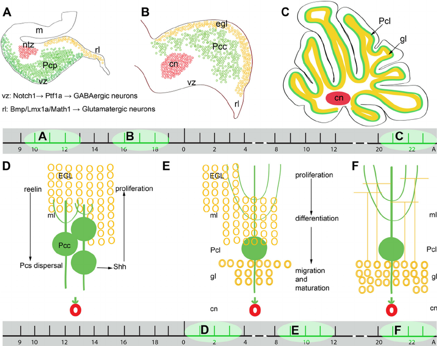
Granule cell (yellow) and purkinje cell (green) migration timetable in mouse cerebellum. Top shows sagittal sections of entire cerebellum. Bottom shows movement of individual cells at interface of EGL and purkinje cell layer.
File history
Click on a date/time to view the file as it appeared at that time.
| Date/Time | Thumbnail | Dimensions | User | Comment | |
|---|---|---|---|---|---|
| current | 10:00, 5 October 2017 | | 850 × 673 (399 KB) | Z5177699 (talk | contribs) | Granule cell (yellow) and purkinje cell (green) migration timetable in mouse cerebellum. Top shows sagittal sections of entire cerebellum. Bottom shows movement of individual cells at interface of EGL and purkinje cell layer. |
You cannot overwrite this file.
File usage
The following 3 pages use this file: