File:Granule Cell and Purkinje Cell Migration.png: Difference between revisions
Original file (850 × 673 pixels, file size: 399 KB, MIME type: image/png)
(No difference)
| |
Latest revision as of 14:09, 16 July 2018
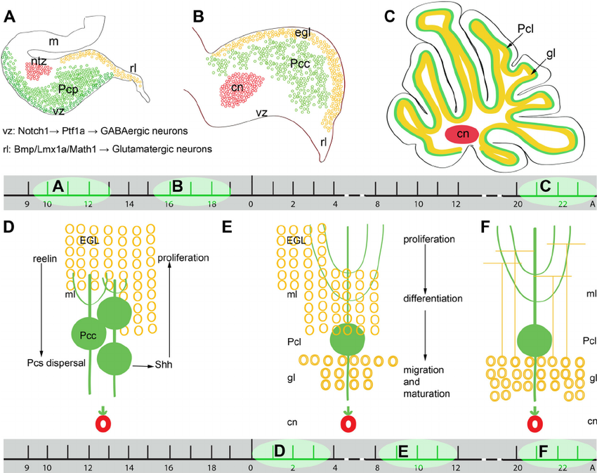
Granule Cell and Purkinje Cell Migration
Granule cell (yellow) and purkinje cell (green) migration timetable in mouse cerebellum.
Top shows sagittal sections of entire cerebellum.
Bottom shows movement of individual cells at interface of EGL and purkinje cell layer.
- Links: cerebellum | neural
Reference
Vriend J, Ghavami S & Marzban H. (2015). The role of the ubiquitin proteasome system in cerebellar development and medulloblastoma. Mol Brain , 8, 64. PMID: 26475605 DOI.
Copyright
© 2015 Vriend et al. Open Access This article is distributed under the terms of the Creative Commons Attribution 4.0 International License (http://creativecommons.org/licenses/by/4.0/), which permits unrestricted use, distribution, and reproduction in any medium, provided you give appropriate credit to the original author(s) and the source, provide a link to the Creative Commons license, and indicate if changes were made. The Creative Commons Public Domain Dedication waiver (http://creativecommons.org/publicdomain/zero/1.0/) applies to the data made available in this article, unless otherwise stated.
- Note - This image was originally uploaded as part of an undergraduate science student project and may contain inaccuracies in either description or acknowledgements. Students have been advised in writing concerning the reuse of content and may accidentally have misunderstood the original terms of use. If image reuse on this non-commercial educational site infringes your existing copyright, please contact the site editor for immediate removal.
File history
Click on a date/time to view the file as it appeared at that time.
| Date/Time | Thumbnail | Dimensions | User | Comment | |
|---|---|---|---|---|---|
| current | 10:00, 5 October 2017 | | 850 × 673 (399 KB) | Z5177699 (talk | contribs) | Granule cell (yellow) and purkinje cell (green) migration timetable in mouse cerebellum. Top shows sagittal sections of entire cerebellum. Bottom shows movement of individual cells at interface of EGL and purkinje cell layer. |
You cannot overwrite this file.
File usage
The following 3 pages use this file: