File:GladstoneWakeley1937 plate03.jpg
Original file (1,827 × 2,504 pixels, file size: 381 KB, MIME type: image/jpeg)
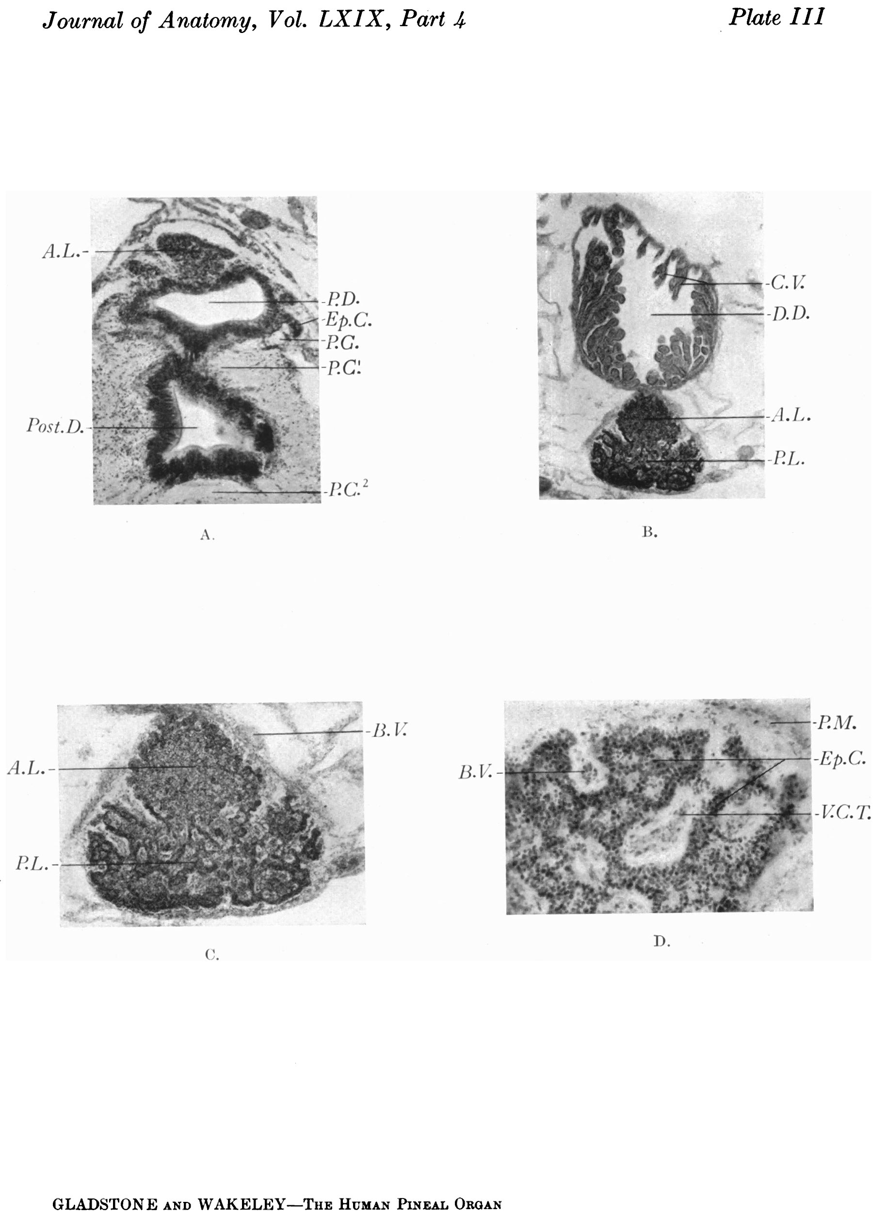
Plate III
Transverse sections of the pineal region of a 6-cm. human embryo, and of a 4.5 months’ foetus.
A. Section through the basal part of the main pineal diverticulum of a 6-cm. human embryo showing, in the upper part of the photograph, the solid anterior lobe. Below this is the main pineal diverticulum, the wall of which shows proliferating cords of ependymal cells growing outward, into the surrounding tissue. Below the pineal evagination is a section through the infrapineal recess, the epithelial lining of which is assuming a columnar type. Fibres of the posterior commissure are seen at the sides and below the pineal region.
B. Coronal section through the pineal region of a 4} months’ human foetus showing the relation of the dorsal diverticulum (suprapineal recess) to the pineal gland. Clusters of elongated chorioidal villi project into the lumen of the diverticulum. The pineal gland shows partial subdivision into an anterior and posterior lobe.
C. The pineal gland more highly magnified showing the ingrowth of vascular processes of the pia mater between the outgrowing neuro-epithelial cords.
D. Peripheral portion of the gland x55 D., showing pale areas containing a central core of vascular pia mater, alternating with dark zones, composed of neuro-epithelial cords.
File history
Click on a date/time to view the file as it appeared at that time.
| Date/Time | Thumbnail | Dimensions | User | Comment | |
|---|---|---|---|---|---|
| current | 10:28, 4 January 2017 | | 1,827 × 2,504 (381 KB) | Z8600021 (talk | contribs) |
You cannot overwrite this file.
File usage
The following page uses this file: