File:GladstoneWakeley1937 plate02.jpg
Original file (1,796 × 2,524 pixels, file size: 632 KB, MIME type: image/jpeg)
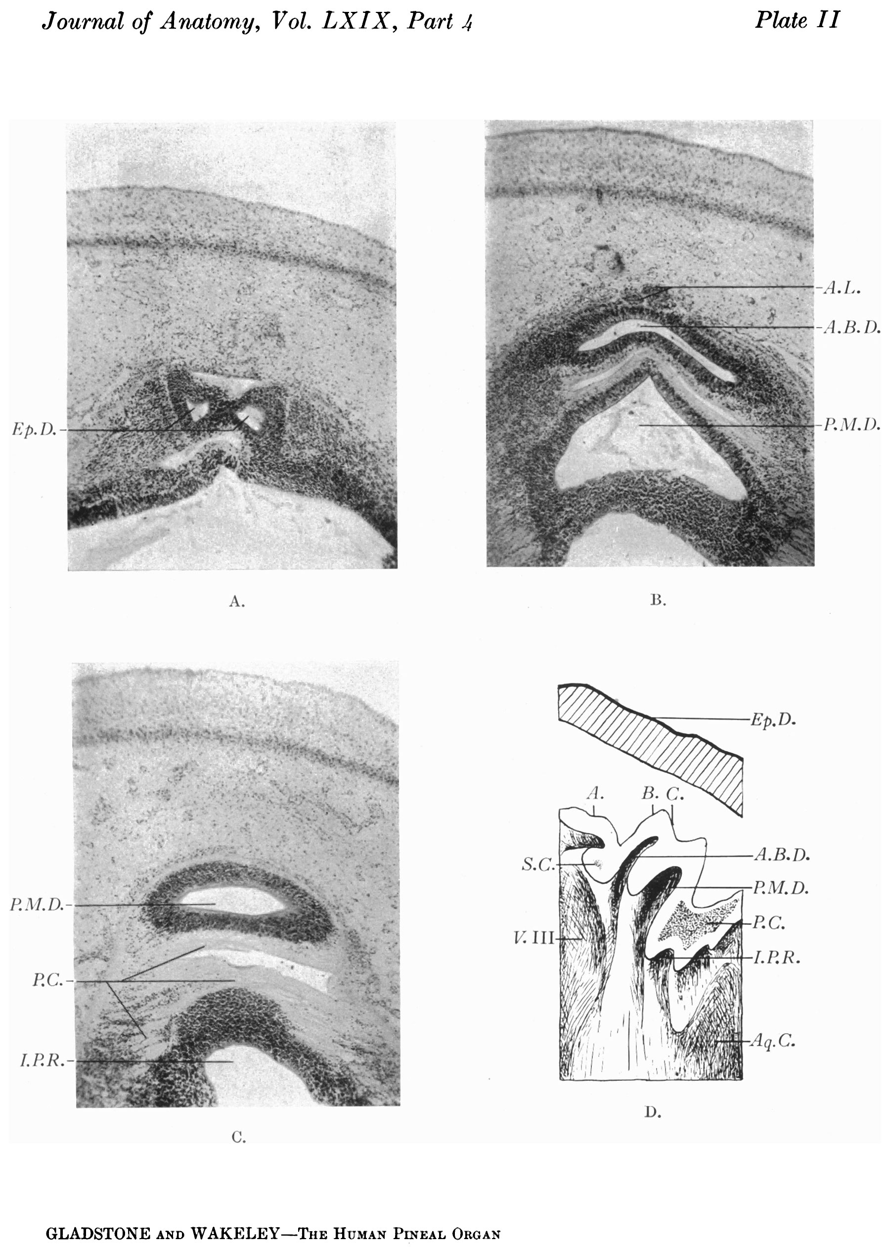
Plate II
Transverse sections of pineal region of a 22-mm. human embryo and median aspect of the right half of a model reconstructed from the corresponding series of sections.
A. Section through the posterior bifurcated end of the anterior ependymal diverticulum, in the plane A of the model.
B. Section through the anterior bilobed diverticulum, and posterior median diverticulum, in the plane B of the model.
C. Section through the posterior median diverticulum and posterior commissure in the plane C of the model.
D. Drawing of the median aspect of the right half of the model showing the relations of the superior and posterior commissures to the pineal diverticula.
| Figure Abbreviations |
|---|
| A.B.D. Anterior bilobed diverticulum.
Ant.D. Anterior diverticulum. A.L. Anterior lobe. Aq.C. Aqueductus cerebri. B. V. Blood vessel. C'. Constriction between “pineal sac” and “pineal eye”. Ca. Capsule. Cap. Capillary. Cav. Cavity. Cbl. Cerebellum. C.C. Corpus callosum. C'.E., C.Ep. Columuar epithelium. C.H. Cerebral hemisphere. Ch.P. Chorioid plexus. C'h.P.V. III Chorioid plexus of third ventricle. C'h.P.L. V. Chorioid plexus of lateral ventricle. C.M. Corpus mammillaris. C.N. III Cranial nerve III. C.N. IV Cranial nerve IV. 0.N. V Cranial nerve V. Cp. Capillary. C.T. Connective tissue. C.T.C’. Connective tissue cells. Cr.C'. Cranial capsule. Cyst. Cyst containing chorioidal villi. C. V. Chorioidal villi. D.D., D.D.’, D.D.” Dorsal diverticulum and its subdivisions. D.S. Dorsal sac. D.M. Dura mater. E.L.M. External limiting membrane. End.S. Endothelial space. Ep. Ependyma. Ep.D. Ependymal diverticulum (dorsal sac). Epd. Epidermis. Ep.Z. Ependymal zone. Ep.C. Epithelial column. Iv'.(.'. Vessels in fibrous capsule. F .F’. F ornix. G.C. V. Great cerebral vein (Galen). G'li., G.S'l2. Glial sheath. Gl.St. Glial stratum (pseudo-epithelium). H.G. Habenular ganglion. I.S.S. Inferior sagittal sinus. I.L.S. Interlobar septum. I.Lr.S. Interlobular septum. Inf. Infundibulum. I.P.R. Infrapineal recess. L. Lobule. Le. Lens. L.N.F.I’.S. Layer of nerve fibres of pineal sac. L.N.F.R. Layer of nerve fibres of retina. Lmn. Lumen. M.B. Meynert’s bundle. M.Z. Marginal zone. N.Z. Nuclear zone. 0p.P.St. Opening of pineal stalk. 0.T. Optic thalamus. P. Pulvinar. Pa. ? Paraphysis. 'Pa.C. Parenchyma cell. Pa.0. Parietal organ (pineal eye). Par. Parenchyma. P.B. Pineal Body. P.O. Pineal cells. P.Co., P.C'., P.0.1, P.C.3 Posterior commissure. P.Cyst. Pineal cyst. P.D. Pineal diverticulum. P.E. Pineal eye (parietal organ). P.I.P. Posterior-intercalary plate. P.L. Posterior lobe. P.M., P.M.1, P.M.’ Pia mater. P.M.D. Posterior median diverticulum. P.0. Pineal organ. Post.D. Posterior diverticulum. P.P.0. Peduncle of pineal organ. P.R. Pineal recess. Pr. Ep. Epithelial process. P.S. Pineal sac. P.Sh. Pial sheath. P.St. Pineal stalk. P. V. Pons Varolii. P. V.A . Post-velar arch. Pv.Sp. Perivascular space. Q.P. Quadrigeminal plate. R. Retina. Ros. “ Rosettes ” . R.P. Rathke’s pouch. S. Septum interhemisphericum. S.C’. Superior commissure (habenular commissure). S.C'.0. Subcommissural organ. S.Co. Sinus confluens. S.0ol. Superior colliculus. S.D. Secondary diverticulum. S.E. condaly evagination. S.D.N. Small darkly stained nucleus. Sp. Space. S.P.L. Splenium. S.T. Sinus transversus. SLO. Stieda’s organ (terminal vesicle). Sub.ep. Subependymal layer. T. Tongue. T.Cbl. Tentorium cerebelli. Th. Thalamus. Tag. Tegmentum. Thr. Thrombus. V.O'.M. Great cerebral vein (V.M. Galeni). V.C.T. Vascular connective tissue. V.F. Velar fold. V.M. Ventriculus mesencephalicus. V.L. Ventriculus lateralis. V. III Third ventricle. V. IV Fourth ventricle. V.S. Venous sinuses. |
| Historic Disclaimer - information about historic embryology pages |
|---|
 |
- Links: fig 1 | fig 2 | fig 3 | fig 4 | fig 5 | fig 6 | fig 7 | fig 8 | fig 9 | fig 10 | fig 18 | plate 1 | plate 2 | plate 3 | plate 4 | plate 5 | plate 6 | 1937 Gladstone Wakeley | Endocrine - Pineal Development | Category:Pineal
Reference
Gladstone RJ. and Wakeley CPG. Development and histogenesis of the human pineal organ. (1937) J. Anat., 19(4): 431-454.
Cite this page: Hill, M.A. (2024, April 25) Embryology GladstoneWakeley1937 plate02.jpg. Retrieved from https://embryology.med.unsw.edu.au/embryology/index.php/File:GladstoneWakeley1937_plate02.jpg
- © Dr Mark Hill 2024, UNSW Embryology ISBN: 978 0 7334 2609 4 - UNSW CRICOS Provider Code No. 00098G
File history
Click on a date/time to view the file as it appeared at that time.
| Date/Time | Thumbnail | Dimensions | User | Comment | |
|---|---|---|---|---|---|
| current | 10:32, 4 January 2017 | | 1,796 × 2,524 (632 KB) | Z8600021 (talk | contribs) |
You cannot overwrite this file.
File usage
The following page uses this file: