File:GladstoneHamilton1941 plate02.jpg

Original file (1,280 × 1,802 pixels, file size: 411 KB, MIME type: image/jpeg)
Plate 2
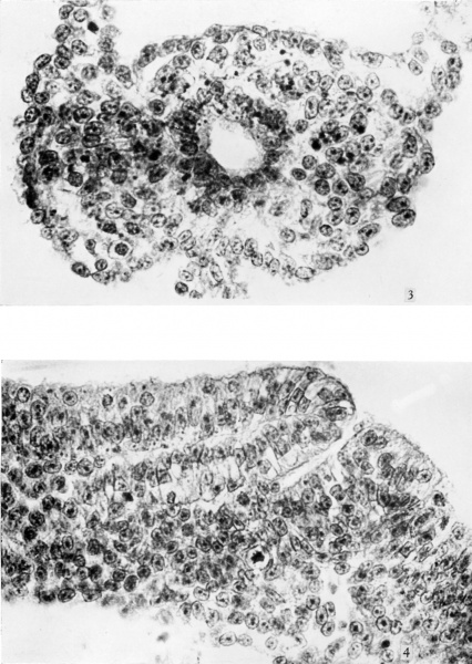
3. A section through the umbilical stalk in the region of the allanto-enteric diverticulum. Chromatic particles are conspicuous in many of the mesodermal cells, and are also present in some of the ectodermal and entodermal cells. Ventro-lateral to the allanto-enteric diverti culum is a group of primitive erythroblasts some of which are undergoing Section no. 108. x 560.
4. Section through the blastopore and beginning of the chorda canal. The anterior lip of the blastopore lies to the left of the photograph. The cells are columnar both in the superficial ectoderm and in the roof of the chorda canal. The nuclei are deeply stained and are, for the most part, situated deeply, near the basement membrane. The cytoplasm is clear and the cell boundaries are mostly well defined. Section no. 46. x 560.
Reference
Gladstone RJ. and Hamilton WJ. A presomite human embryo (Shaw) with primitive streak and chorda canal with special reference to the development of the vascular system. (1941) Amer. J Anat. 76(1): 9-44.
Cite this page: Hill, M.A. (2024, April 25) Embryology GladstoneHamilton1941 plate02.jpg. Retrieved from https://embryology.med.unsw.edu.au/embryology/index.php/File:GladstoneHamilton1941_plate02.jpg
- © Dr Mark Hill 2024, UNSW Embryology ISBN: 978 0 7334 2609 4 - UNSW CRICOS Provider Code No. 00098G
File history
Click on a date/time to view the file as it appeared at that time.
| Date/Time | Thumbnail | Dimensions | User | Comment | |
|---|---|---|---|---|---|
| current | 16:50, 26 February 2017 |  | 1,280 × 1,802 (411 KB) | Z8600021 (talk | contribs) | |
| 16:50, 26 February 2017 |  | 1,586 × 2,521 (916 KB) | Z8600021 (talk | contribs) |
You cannot overwrite this file.