File:Gastrointestinal tract intestine immune cartoon 01.jpg
Original file (728 × 1,200 pixels, file size: 284 KB, MIME type: image/jpeg)
Gastrointestinal Tract Immune
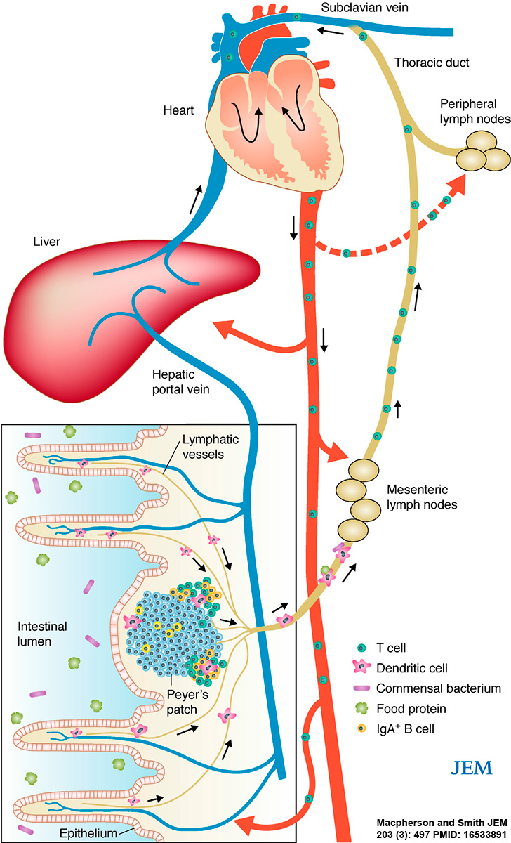
This cartoon gives an overview of the intestinal immune system interaction through Mesenteric Lymph Nodes (MNLs) with the circulating immune system.
Functional anatomy of induction of immune responses by intestinal antigens. Abundant protein antigens and live commensal bacteria are present in the intestine. Antigenic peptides can pass into the bloodstream through one of the tributaries of the hepatic portal vein or are taken up by DCs in the subepithelial region of the Peyer's patches and carried to the MLNs via the afferent lymphatics. Although it is possible for circulating peptides to tolerize T cells in the liver or peripheral lymph nodes, presentation in the MLNs is the dominant tolerogenic pathway. Commensal bacteria are also sampled by intestinal DCs and induce IgA responses in the Peyer's patches; although very small numbers of commensals can be carried to MLN by DC, systemic tolerance to these organisms is not induced. Because the commensal laden DCs do not penetrate further than the MLN, the systemic immune system is protected from unwanted priming reactions from live bacteria.
- Immune Images: Oesophagus MALT | Colon MALT | Peyer's patch overview | Peyer's patch detail | Cartoon - IEL development | Cartoon - IEL function | Cartoon - IEL differentiation | Mesenteric Lymph Nodes overview | Palatine Tonsil | Tonsil | Immune System Development
Reference
<pubmed>16533891</pubmed>| PMC2118258 | J Exp Med.
Published March 20, 2006 // JEM vol. 203 no. 3 497-500 The Rockefeller University Press, doi: 10.1084/jem.20060227
Copyright
Rockefeller University Press - Copyright Policy This article is distributed under the terms of an Attribution–Noncommercial–Share Alike–No Mirror Sites license for the first six months after the publication date (see http://www.jcb.org/misc/terms.shtml). After six months it is available under a Creative Commons License (Attribution–Noncommercial–Share Alike 4.0 Unported license, as described at https://creativecommons.org/licenses/by-nc-sa/4.0/ ). (More? Help:Copyright Tutorial)
Figure 2. modified in size and labelling. Text above modified from figure legend.
Cite this page: Hill, M.A. (2024, April 19) Embryology Gastrointestinal tract intestine immune cartoon 01.jpg. Retrieved from https://embryology.med.unsw.edu.au/embryology/index.php/File:Gastrointestinal_tract_intestine_immune_cartoon_01.jpg
- © Dr Mark Hill 2024, UNSW Embryology ISBN: 978 0 7334 2609 4 - UNSW CRICOS Provider Code No. 00098G
File history
Click on a date/time to view the file as it appeared at that time.
| Date/Time | Thumbnail | Dimensions | User | Comment | |
|---|---|---|---|---|---|
| current | 10:31, 30 January 2015 | | 728 × 1,200 (284 KB) | Z8600021 (talk | contribs) | ==Gastrointestinal tract immune== Functional anatomy of induction of immune responses by intestinal antigens. Abundant protein antigens and live commensal bacteria are present in the intestine. Antigenic peptides can pass into the bloodstream through... |
You cannot overwrite this file.
File usage
The following 4 pages use this file: