File:Gaba-effects-retina.JPG
Original file (879 × 2,084 pixels, file size: 240 KB, MIME type: image/jpeg)
"GABAA receptor mediated effects on retinal progenitor cell proliferation."
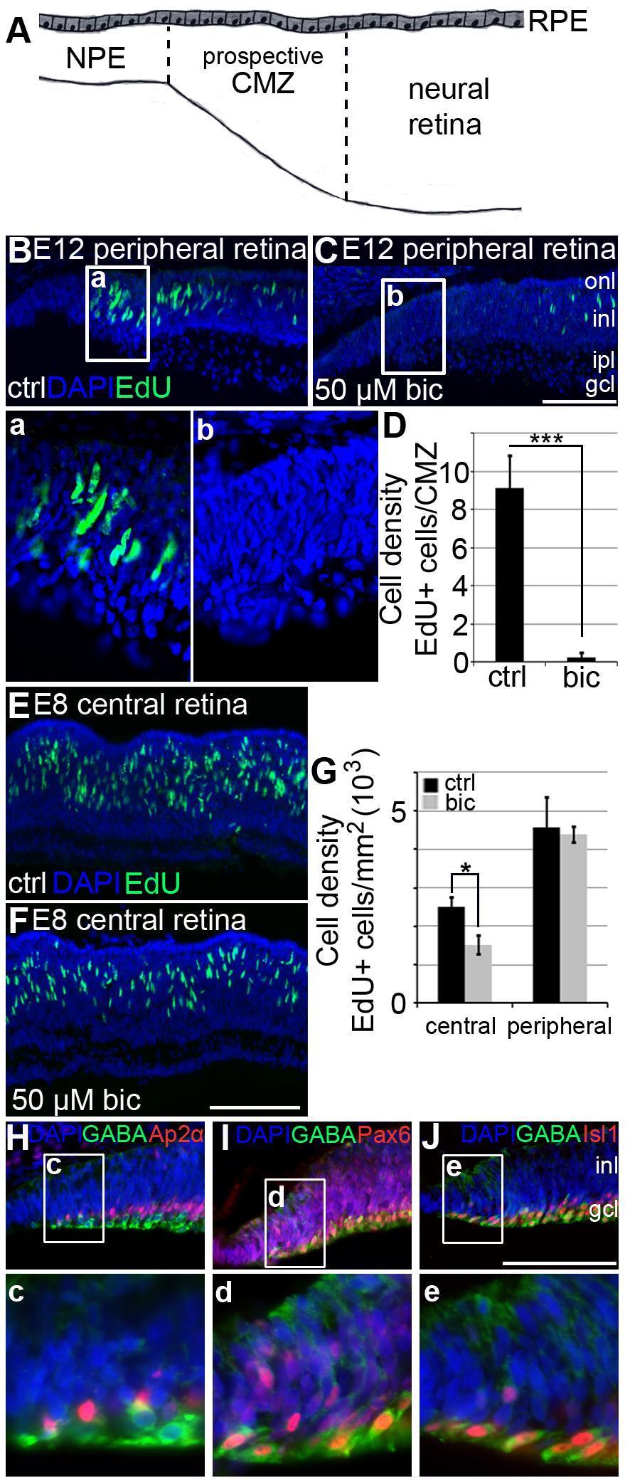
"(A) Schematic diagram of the E12 peripheral retina. Cells between the dashed lines in the prospective ciliary marginal zone (CMZ) on the dorsal side of the retina were counted. Cells that had gone through S-phase were detected by EdU labelling. Fluorescence micrographs of EdU labelled cells in (B) untreated (control) and (C) bicuculline-treated E12 retinal explants cultured for 4 hours. Images in (a) and (b) show the boxed regions in (B) and (C). (D) Bar graph shows the cell density of EdU positive cells per CMZ in control and bicuculline-treated explants. Error bars ± S.D. n = 4 explant cultures, average of 4 sections per explant, two-tailed Student t-test; *** p<0.001. Fluorescence micrographs of EdU labelled cells in the central retina in (E) control and (F) bicuculline-treated E8 explants cultured for 4 hours. (G) Bar graph shows cell density of EdU positive cells in central and peripheral retina in control (black bars) and bicuculline-treated (grey bars) E8 explants. Error bars ± S.D. n = 3 explants cultures, average of 4 sections per explant, Mann-Whitney test; * p = 0.05. (H–J) GABA+ cells close and within the prospective CMZ labelled for (H) Ap2α, (I) Pax6 or (J) Isl1. Images in (c) to (e) show the boxed regions in (H–J) in higher magnification. gcl, ganglion cell layer; inl, inner nuclear layer; ipl, inner plexiform layer; onl, outer nuclear layer; RPE, retinal pigment epithelium. Scale bar in (C) is 100 µm and is valid for (B). Scale bar in (F) is 100 µm and is valid for (E). Scale bar in (J) is 100 µm is valid for (H–J)."
doi:10.1371/journal.pone.0036874.g003
Source: <pubmed>22590629</pubmed>
Copyright information: © 2012 Ring et al. This is an open-access article distributed under the terms of the Creative Commons Attribution License, which permits unrestricted use, distribution, and reproduction in any medium, provided the original author and source are credited.
- Note - This image was originally uploaded as part of an undergraduate science student project and may contain inaccuracies in either description or acknowledgements. Students have been advised in writing concerning the reuse of content and may accidentally have misunderstood the original terms of use. If image reuse on this non-commercial educational site infringes your existing copyright, please contact the site editor for immediate removal.
File history
Click on a date/time to view the file as it appeared at that time.
| Date/Time | Thumbnail | Dimensions | User | Comment | |
|---|---|---|---|---|---|
| current | 20:43, 2 October 2012 | | 879 × 2,084 (240 KB) | Z3370664 (talk | contribs) | "GABAA receptor mediated effects on retinal progenitor cell proliferation." "(A) Schematic diagram of the E12 peripheral retina. Cells between the dashed lines in the prospective ciliary marginal zone (CMZ) on the dorsal side of the retina were counted. |
You cannot overwrite this file.
File usage
The following page uses this file: