File:Frazer1926 fig03.jpg: Difference between revisions
mNo edit summary |
mNo edit summary |
||
| Line 7: | Line 7: | ||
{{Frazer1926 figures}} | {{Frazer1926 figures}} | ||
][[Category:Carnegie Stage 13]][[Category:Week 4]][[Category:Week 5]] | |||
Latest revision as of 15:04, 29 July 2015
Fig. 3. Head of Embryo (8 mm)
The precervical sinus in an embryo of 8 mm. seen from the left side. For description see text.
At the point ep. in fig. 3, where the growing paraxial region is apposed to the long second arch, there is a very small fusion between these parts on the other side in this embryo; it could not be recognised with certainty on the left side. It suggests a fusion, no doubt associated with the growth which causes the overlapping of the placode here, between the paraxial swelling and the upper part of the hinder border of the second arch. The upper portion of the second groove, above the level of the placode, is now only represented by a shallow depression. This part has been practically obliterated, but not, it would appear, by any process of fusion: the condition found in an embryo of 7 mm. supports this opinion. In this specimen the paraxial growth is overhanging the placode but has not yet begun to turn it in, and the posterior border of the second arch, clearly cut opposite the placode, becomes shallow and indefinite above this in the region of paraxial thickening. It seems that the level of the depression behind the upper part of the second arch, as far down as the placodal inturning, is simply raised and the edge of the arch made indefinite by the growth of the mass behind it, without any meeting and fusion of the two prominences. If this view of the matter is correct, the very small fusion noticed on one side in the 8mm. embryo may be nothing but a deceptive appearance, or may be an indication of a definite little point of suture between the arch and the overhanging lip of the growth covering in the placodal area. The fusion, however, if it does occur at all, does not go any further: the conditions shown in fig.3, in fact, may be said to indicate already what will be the extent of overlapping in the region of the sinus brought about by the surrounding growth.
| Historic Disclaimer - information about historic embryology pages |
|---|
 |
- Links: Fig. 1. Embryo 4-9 mm | Fig. 2. Semi-schematic pharyngeal region | Fig. 3. Embryo 8 mm | Fig.4. Embryo 10 mm | Fig. 5. Embryo 12 mm | Fig. 6. Embryo 10 mm | Fig. 7. Third Arch | Fig. 8. Laranryngeal Area of Head | Plate 1. Fig.1,3,4
Reference
Frazer JE. The disappearance of the precervical sinus. (1926) J Anat. 61(1): 132-43. PMID 17104123.
Cite this page: Hill, M.A. (2024, April 19) Embryology Frazer1926 fig03.jpg. Retrieved from https://embryology.med.unsw.edu.au/embryology/index.php/File:Frazer1926_fig03.jpg
- © Dr Mark Hill 2024, UNSW Embryology ISBN: 978 0 7334 2609 4 - UNSW CRICOS Provider Code No. 00098G
]
File history
Click on a date/time to view the file as it appeared at that time.
| Date/Time | Thumbnail | Dimensions | User | Comment | |
|---|---|---|---|---|---|
| current | 10:42, 28 July 2015 | 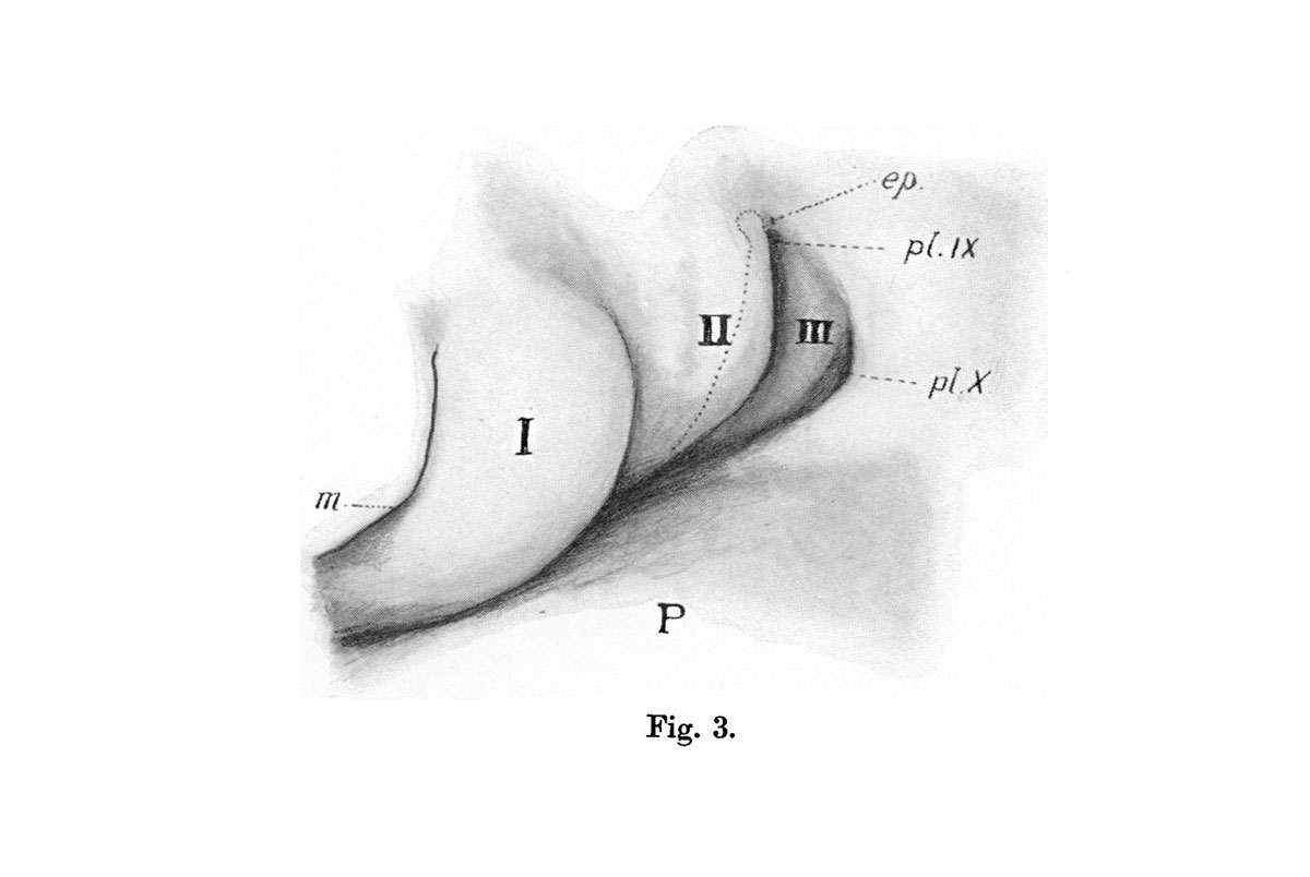 | 1,200 × 804 (69 KB) | Z8600021 (talk | contribs) |
You cannot overwrite this file.
File usage
The following 2 pages use this file: