File:Frazer1926 fig02.jpg: Difference between revisions
From Embryology
(Z8600021 uploaded a new version of File:Frazer1926 fig02.jpg) |
mNo edit summary |
||
| Line 1: | Line 1: | ||
==Fig. 2== | ==Fig. 2. Semi-schematic view of the lower aspect of the pharyngeal region== | ||
The pericardium having been removed, save for its roof. I, II, III, IV, pharyngeal arches, external; P, roof of pericardium. The whole region is somewhat flattened out to show the relations. substance of the mandibular arch, which is the only one completely joined in the middle line. | |||
{{Frazer1926 figures}} | {{Frazer1926 figures}} | ||
Revision as of 10:57, 28 July 2015
Fig. 2. Semi-schematic view of the lower aspect of the pharyngeal region
The pericardium having been removed, save for its roof. I, II, III, IV, pharyngeal arches, external; P, roof of pericardium. The whole region is somewhat flattened out to show the relations. substance of the mandibular arch, which is the only one completely joined in the middle line.
| Historic Disclaimer - information about historic embryology pages |
|---|
 |
- Links: Fig. 1. Embryo 4-9 mm | Fig. 2. Semi-schematic pharyngeal region | Fig. 3. Embryo 8 mm | Fig.4. Embryo 10 mm | Fig. 5. Embryo 12 mm | Fig. 6. Embryo 10 mm | Fig. 7. Third Arch | Fig. 8. Laranryngeal Area of Head | Plate 1. Fig.1,3,4
Reference
Frazer JE. The disappearance of the precervical sinus. (1926) J Anat. 61(1): 132-43. PMID 17104123.
Cite this page: Hill, M.A. (2024, April 18) Embryology Frazer1926 fig02.jpg. Retrieved from https://embryology.med.unsw.edu.au/embryology/index.php/File:Frazer1926_fig02.jpg
- © Dr Mark Hill 2024, UNSW Embryology ISBN: 978 0 7334 2609 4 - UNSW CRICOS Provider Code No. 00098G
File history
Click on a date/time to view the file as it appeared at that time.
| Date/Time | Thumbnail | Dimensions | User | Comment | |
|---|---|---|---|---|---|
| current | 10:56, 28 July 2015 | 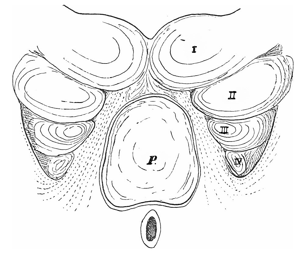 | 991 × 833 (145 KB) | Z8600021 (talk | contribs) | |
| 10:56, 28 July 2015 | 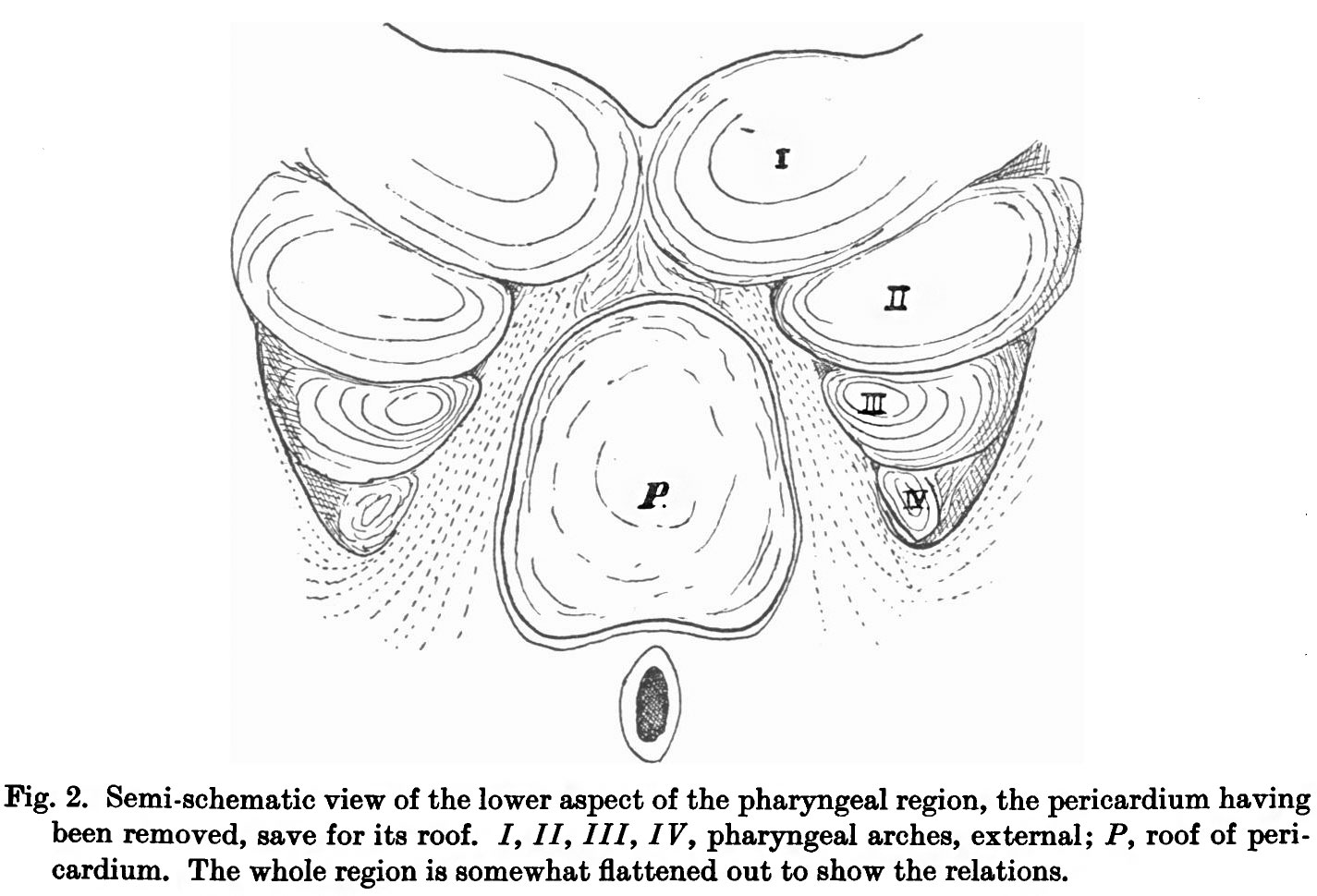 | 1,425 × 970 (180 KB) | Z8600021 (talk | contribs) |
You cannot overwrite this file.
File usage
The following page uses this file: