File:Frazer1921 fig01.jpg

Original file (974 × 1,200 pixels, file size: 104 KB, MIME type: image/jpeg)
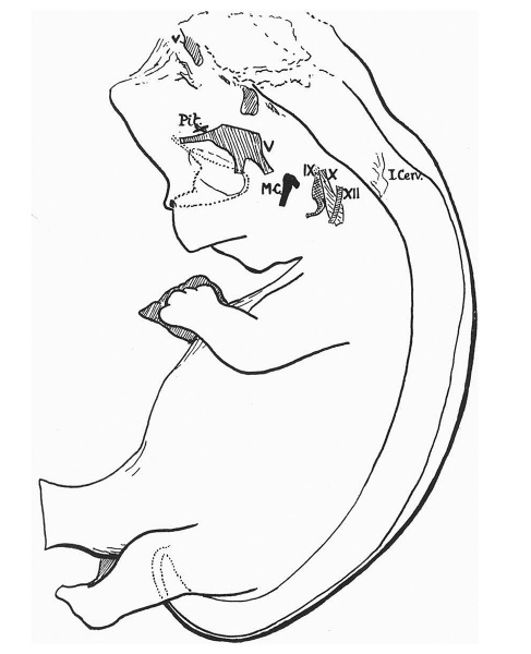
Fig. 1. Linear reconstruction of anencephalic embryo
The shaded areas in the head indicate certain nerve positions. V, the fifth nerve and its attached root; IX, X, XII are corresponding nerves. Some nerve remnants lie along the ventral surface of the elongated hind-brain stem. Pit,., marks the situation of the pituitary rudiment. M.C. is the upper end of Meckel’s cartilage.
| Historic Disclaimer - information about historic embryology pages |
|---|
 |
Reference
Frazer JE. Report on an Anencephalic Embryo PMID 17103933
Cite this page: Hill, M.A. (2024, April 25) Embryology Frazer1921 fig01.jpg. Retrieved from https://embryology.med.unsw.edu.au/embryology/index.php/File:Frazer1921_fig01.jpg
- © Dr Mark Hill 2024, UNSW Embryology ISBN: 978 0 7334 2609 4 - UNSW CRICOS Provider Code No. 00098G
File history
Click on a date/time to view the file as it appeared at that time.
| Date/Time | Thumbnail | Dimensions | User | Comment | |
|---|---|---|---|---|---|
| current | 17:55, 15 February 2016 | | 974 × 1,200 (104 KB) | Z8600021 (talk | contribs) | |
| 17:53, 15 February 2016 | 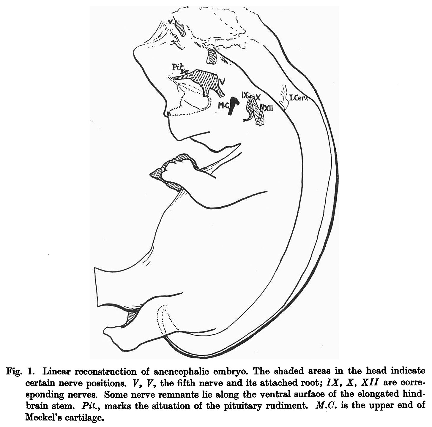 | 1,454 × 1,438 (209 KB) | Z8600021 (talk | contribs) |
You cannot overwrite this file.
File usage
The following 2 pages use this file: