File:Fragile site appearance and distribution.png
Fragile_site_appearance_and_distribution.png (548 × 520 pixels, file size: 101 KB, MIME type: image/png)
Screen_shot_2011-08-18_at_8.47.56_AM.png
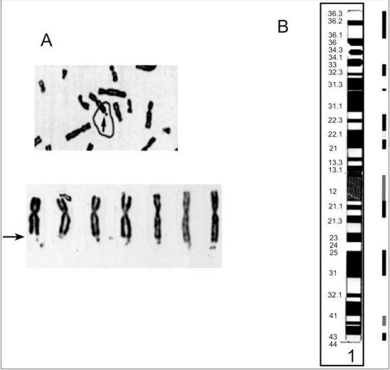
Fig. (7) Fragile site appearance and distribution. A. Cytogenetic appearance of fragile X. Arrows point to fragile sites. B. Distributation of fragile sites along chromosome 1. The bars beside the cytogenetic bands represent the fragile site locations (see Table 11). Dark to light bars represent inducing agents. Amphidicolin, 5-Azacytidine, and Folic acid, respectively. Taken from [150].
http://www.ncbi.nlm.nih.gov/pmc/articles/PMC3018726/
This is an open access article distributed under the terms of the Creative Commons Attribution License (http://creativecommons.org/licenses/by/2.5/), which permits unrestrictive use, distribution, and reproduction in any medium, provided the original work is properly cited.
File history
Click on a date/time to view the file as it appeared at that time.
| Date/Time | Thumbnail | Dimensions | User | Comment | |
|---|---|---|---|---|---|
| current | 08:51, 18 August 2011 | | 548 × 520 (101 KB) | Z3290618 (talk | contribs) | Screen_shot_2011-08-18_at_8.47.56_AM.png Fig. (7) Fragile site appearance and distribution. A. Cytogenetic appearance of fragile X. Arrows point to fragile sites. B. Distributation of fragile sites along chromosome 1. The bars beside the cytogenetic band |
You cannot overwrite this file.
File usage
The following page uses this file: