File:Foster006.jpg
Original file (1,034 × 515 pixels, file size: 72 KB, MIME type: image/jpeg)
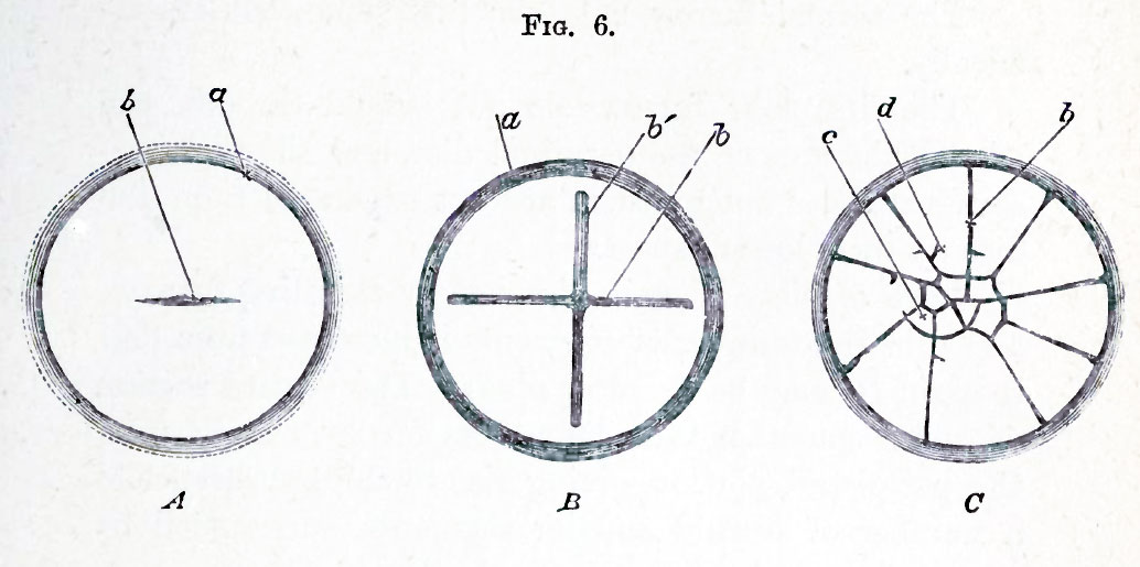
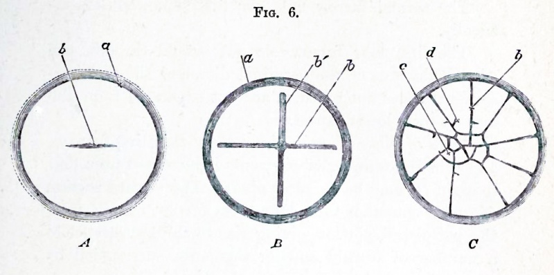
FIG. 6. SURFACE VIEWS OF THE EARLY STAGES OF THE SEGMENTATION IN A FOWL'S EGG. (A and G after Coste.)
A represents the earliest stage. The first furrow (b) has begun to make its appearance in the centre of the germinal disc, whose periphery is marked by the line a. In J3 } the first furrow is completed nearly across the disc, and a second similar furrow at right angles to the first has appeared. The disc thus becomes divided somewhat irregularly into quadrants by four (half) furrows. In a later stage ((7) the meridian furrows b have increased in number, from four, as in B, to nine, and cross furrows have also made their appearance. The disc is thus cut up into small central (c) and larger peripheral (d) segments. Several new cross furrows are seen just beginning, as ex. gr. close to the end of the line of reference d.
Viewed from above, a furrow is seen to make its appearance, running across the germinal disc, though not for the whole breadth, and dividing it into two halves (Fig. 6, A). This primary furrow is succeeded by a second at right angles to itself. The surface thus becomes divided into four segments or quadrants (Fig. 6, B).
| Historic Disclaimer - information about historic embryology pages |
|---|
 |
Reference
Foster, M., Balfour, F. M., Sedgwick, A., & Heape, W. (1883). The Elements of Embryology. (2nd ed.). London: Macmillan and Co.
The Elements of Embryology (1883)
File history
Click on a date/time to view the file as it appeared at that time.
| Date/Time | Thumbnail | Dimensions | User | Comment | |
|---|---|---|---|---|---|
| current | 15:34, 8 January 2011 | | 1,034 × 515 (72 KB) | S8600021 (talk | contribs) | FIG. 6. SURFACE VIEWS OF THE EARLY STAGES OF THE SEGMENTATION IN A FOWL'S EGG. (A and G after Coste.) A represents the earliest stage. The first furrow (b) has begun to make its appearance in the centre of the germinal disc, whose periphery is marked by |
You cannot overwrite this file.
File usage
The following 2 pages use this file: