File:Foster004.jpg: Difference between revisions
(FIG. 4. SECTION THROUGH THE GERMINAL Disc OF THE RIPE OVARIAN OVUM OF A FOWL WHILE YET ENCLOSED IN ITS CAPSULE. a. Connective-tissue capsule of the ovum. b. follicular epithelium, at the surface of which nearest the ovum lies the vitelline membrane, c) |
No edit summary |
||
| (One intermediate revision by one other user not shown) | |||
| Line 1: | Line 1: | ||
==Fig. 4. Section through the germinal disc of the ripe ovarian ovum of a fowl while yet enclosed in its capsule== | |||
a. Connective-tissue capsule of the ovum. | a. Connective-tissue capsule of the ovum. | ||
| Line 14: | Line 15: | ||
{{Template:Foster 1883 Figures}} | |||
{{Template:Foster 1883 | |||
Latest revision as of 11:08, 22 December 2012
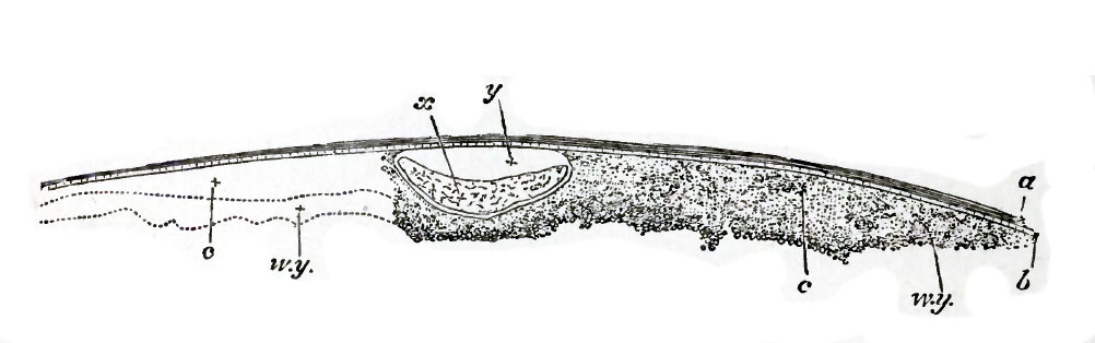
Fig. 4. Section through the germinal disc of the ripe ovarian ovum of a fowl while yet enclosed in its capsule
a. Connective-tissue capsule of the ovum.
b. follicular epithelium, at the surface of which nearest the ovum lies the vitelline membrane,
c. granular material of the germinal disc, which becomes converted into the blastoderm. (This is not very well represented in the woodcut. In sections which have been hardened in chromic acid it consists of fine granules.)
w.y, white yolk, which passes insensibly into the fine granular material of the disc.
x, germinal vesicle enclosed in a distinct membrane, but shrivelled up by the action of the chromic acid,
y, space originally completely filled up by the germinal vesicle, before the latter was shrivelled up by the action of the chromic acid.
| Historic Disclaimer - information about historic embryology pages |
|---|
 |
Reference
Foster, M., Balfour, F. M., Sedgwick, A., & Heape, W. (1883). The Elements of Embryology. (2nd ed.). London: Macmillan and Co.
The Elements of Embryology (1883)
File history
Click on a date/time to view the file as it appeared at that time.
| Date/Time | Thumbnail | Dimensions | User | Comment | |
|---|---|---|---|---|---|
| current | 14:59, 8 January 2011 | 1,002 × 314 (45 KB) | S8600021 (talk | contribs) | FIG. 4. SECTION THROUGH THE GERMINAL Disc OF THE RIPE OVARIAN OVUM OF A FOWL WHILE YET ENCLOSED IN ITS CAPSULE. a. Connective-tissue capsule of the ovum. b. follicular epithelium, at the surface of which nearest the ovum lies the vitelline membrane, c |
You cannot overwrite this file.
File usage
The following 2 pages use this file: