File:Finley1923 fig06.jpg: Difference between revisions
No edit summary |
No edit summary |
||
| (One intermediate revision by the same user not shown) | |||
| Line 1: | Line 1: | ||
==Figure 6 Photograph from a total mount of a piece of the scalp from a human embryo 28 mm in length== | |||
Photograph from another portion of the same section as above, showing, under higher magnification, the first and second zones. In the center a long tip from the angioblastic plexus is seen to penetrate the avascular zone. This represents the first step in the differentiation of the mesenchyme into angioblastic tissue. X 150. | |||
Photograph from a total mount of a piece of the scalp from a human embryo 28 mm. in length (No. 1240a). | |||
{{Finley1923}} | |||
Latest revision as of 16:04, 12 August 2012
Figure 6 Photograph from a total mount of a piece of the scalp from a human embryo 28 mm in length
Photograph from another portion of the same section as above, showing, under higher magnification, the first and second zones. In the center a long tip from the angioblastic plexus is seen to penetrate the avascular zone. This represents the first step in the differentiation of the mesenchyme into angioblastic tissue. X 150.
Photograph from a total mount of a piece of the scalp from a human embryo 28 mm. in length (No. 1240a).
- 1923 Head Subcutaneous Plexus: Plate 1 | Plate 2 | Fig 1 | Fig 2 | Fig 3 | Fig 4 | Fig 5 | Fig 6 | Fig 7 | Fig 8 | Fig 9 | Fig 10 | Fig 11 | Fig 12 | Fig 13 | Carnegie No.71 | Historic Disclaimer
| Historic Disclaimer - information about historic embryology pages |
|---|
 |
Reference
Finley EB. The development of the subcutaneous vascular plexus in the head of the human embryo. (1923) Contributions to Embryology Carnegie Institution No. 71: 155-161.
Cite this page: Hill, M.A. (2024, April 19) Embryology Finley1923 fig06.jpg. Retrieved from https://embryology.med.unsw.edu.au/embryology/index.php/File:Finley1923_fig06.jpg
- © Dr Mark Hill 2024, UNSW Embryology ISBN: 978 0 7334 2609 4 - UNSW CRICOS Provider Code No. 00098G
File history
Click on a date/time to view the file as it appeared at that time.
| Date/Time | Thumbnail | Dimensions | User | Comment | |
|---|---|---|---|---|---|
| current | 16:00, 12 August 2012 | 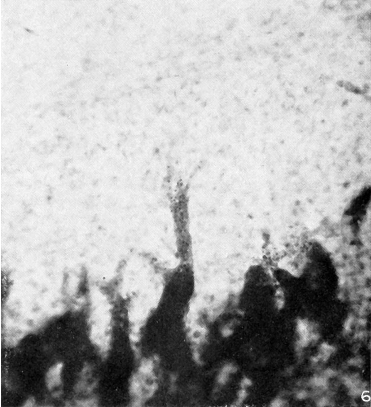 | 729 × 800 (95 KB) | Z8600021 (talk | contribs) |
You cannot overwrite this file.