File:Figure 1 Morphological defects in CTCF mutant embryonic hearts.PNG: Difference between revisions
No edit summary |
m (→Reference) |
||
| (5 intermediate revisions by 2 users not shown) | |||
| Line 1: | Line 1: | ||
===Description=== | ===Description=== | ||
Morphological defects in Ctcf mutant embryonic hearts. | |||
Whole mount control and mutant (Ctcffl/fl;Nkx2-5-Cre) embryos at E10.5 (A, B), E11.5 (C, D) and E12.5 (E, F). Arrows in (E) point to the pericardial edema present in mutant embryos. Higher magnification of the heart show morphological defects in Ctcf mutant hearts becoming manifest at E12.5 (F). (G-L) Sections from control and mutant embryos stained with hematoxylin and eosin; higher magnifications (black dashed boxes) for each are shown on the right of each section. Disorganization of the interventricular septum and thinning of the myocardial wall is apparent in Ctcf mutant hearts at E11.5. A, atria; V, ventricle; RA, right atria; LA, left atria; RV, right ventricle; LV, left ventricle; AVC, atrioventricular canal; IVS, interventricular septum. Scale bars, 750 μm (A), 200 μm (B, D), 1 mm (C), 2 mm (E), 800 μm (F), 100 μm (G, H), 200 μm (I-L), and 100 μm in all higher magnifications. | |||
===Reference=== | ===Reference=== | ||
{{#pmid:28846746}} | |||
===Copyright=== | ===Copyright=== | ||
© 2017 Gomez-Velazquez et al. This is an open access article distributed under the terms of the Creative Commons Attribution License, which permits unrestricted use, distribution, and reproduction in any medium, provided the original author and source are credited. | |||
{{Template:Student Image}} | {{Template:Student Image}} | ||
Latest revision as of 10:46, 18 December 2018
Description
Morphological defects in Ctcf mutant embryonic hearts. Whole mount control and mutant (Ctcffl/fl;Nkx2-5-Cre) embryos at E10.5 (A, B), E11.5 (C, D) and E12.5 (E, F). Arrows in (E) point to the pericardial edema present in mutant embryos. Higher magnification of the heart show morphological defects in Ctcf mutant hearts becoming manifest at E12.5 (F). (G-L) Sections from control and mutant embryos stained with hematoxylin and eosin; higher magnifications (black dashed boxes) for each are shown on the right of each section. Disorganization of the interventricular septum and thinning of the myocardial wall is apparent in Ctcf mutant hearts at E11.5. A, atria; V, ventricle; RA, right atria; LA, left atria; RV, right ventricle; LV, left ventricle; AVC, atrioventricular canal; IVS, interventricular septum. Scale bars, 750 μm (A), 200 μm (B, D), 1 mm (C), 2 mm (E), 800 μm (F), 100 μm (G, H), 200 μm (I-L), and 100 μm in all higher magnifications.
Reference
Gomez-Velazquez M, Badia-Careaga C, Lechuga-Vieco AV, Nieto-Arellano R, Tena JJ, Rollan I, Alvarez A, Torroja C, Caceres EF, Roy AR, Galjart N, Delgado-Olguin P, Sanchez-Cabo F, Enriquez JA, Gomez-Skarmeta JL & Manzanares M. (2017). CTCF counter-regulates cardiomyocyte development and maturation programs in the embryonic heart. PLoS Genet. , 13, e1006985. PMID: 28846746 DOI.
Copyright
© 2017 Gomez-Velazquez et al. This is an open access article distributed under the terms of the Creative Commons Attribution License, which permits unrestricted use, distribution, and reproduction in any medium, provided the original author and source are credited.
- Note - This image was originally uploaded as part of an undergraduate science student project and may contain inaccuracies in either description or acknowledgements. Students have been advised in writing concerning the reuse of content and may accidentally have misunderstood the original terms of use. If image reuse on this non-commercial educational site infringes your existing copyright, please contact the site editor for immediate removal.
File history
Click on a date/time to view the file as it appeared at that time.
| Date/Time | Thumbnail | Dimensions | User | Comment | |
|---|---|---|---|---|---|
| current | 19:07, 3 October 2017 | 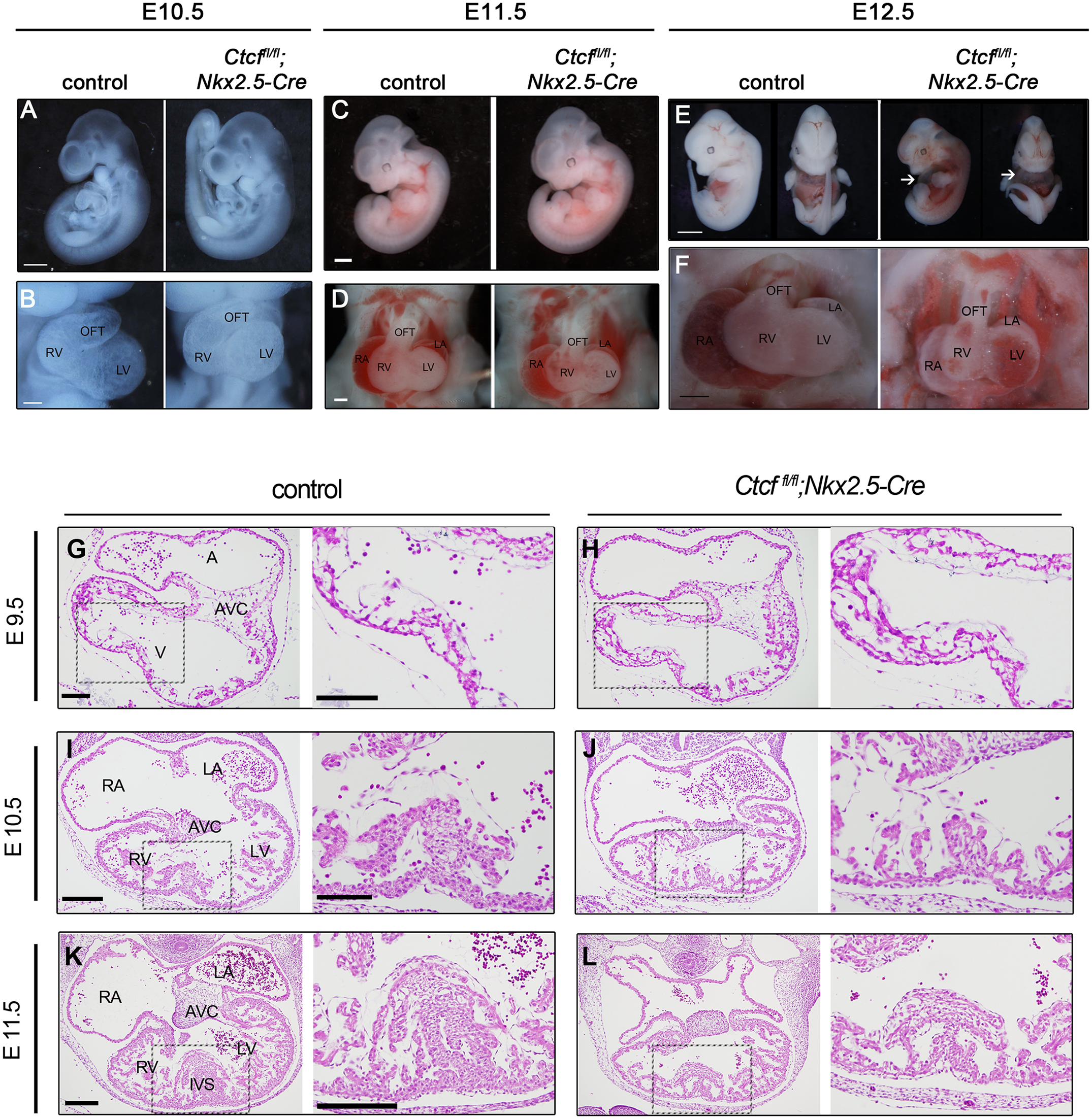 | 2,150 × 2,205 (5.9 MB) | Z5018962 (talk | contribs) |
You cannot overwrite this file.
File usage
The following 2 pages use this file: