File:Fetal head section 01.jpg: Difference between revisions
From Embryology
mNo edit summary |
|||
| (22 intermediate revisions by the same user not shown) | |||
| Line 1: | Line 1: | ||
==Human Fetal Head Week 12== | ==Human Fetal Head Week 12== | ||
This mid-line section through the early fetal head (12 weeks) shows features of the developing {{skull}}, {{neural}} (brain), face, mouth, pharynx and neck. | |||
{ | {| | ||
|-bgcolor="CEDFF2" | |||
! Neural System | |||
! Endocrine | |||
== | ! Skeletal System | ||
* | ! Muscle | ||
* | |- | ||
* lamina terminalis | | valign=top width=200px| | ||
* fourth ventricle | * [[Neural_System_Development|central nervous system]]. | ||
* {{hypothalamus}} | |||
* {{lamina terminalis}} (site of anterior neuropore closure). | |||
* [[Neural_-_Ventricular_System_Development|fourth ventricle]], ventricular space behind the pons and medulla. | |||
* brainstem - {{pons}} and {{medulla oblongata}} | |||
* basilar artery - lying ventral to the pons | * basilar artery - lying ventral to the pons | ||
| valign=top width=200px| | |||
* {{hypothalamus}} | |||
* {{pituitary}} sitting in the sella turcica. | |||
** note remnant of Rathke's pouch still visible on floor of developing sella turcica (cartilage) | |||
| valign=top width=200px| | |||
* {{cartilage}} - {{skull}} septum of the nose. | |||
* {{bone}} - [[Respiratory_System_-_Upper_Respiratory_Tract|upper respiratory]] ossifying nasal concha. | |||
* {{bone}} - [[Palate_Development|hard palate]] roof of mouth. | |||
* {{cartilage}} - base of skull and vertebra. | |||
* {{bone}} - {{mandible}}. | |||
* {{axial skeleton}} - vertebra, axis and atlas. | |||
| valign=top width=200px| | |||
* [[Musculoskeletal_System_-_Muscle_Development|muscle]] - {{tongue}}, attached to mandible, note foramen cecum. | |||
* {{muscle}} - soft {{palate}} back of mouth. | |||
** genioglossus muscle - attached to mandible | |||
** geniohyoideus muscle - attaching hyoid bone to mandible | |||
|} | |||
{{Fetal head 12 week}} | |||
Latest revision as of 09:15, 20 February 2019
Human Fetal Head Week 12
This mid-line section through the early fetal head (12 weeks) shows features of the developing skull, neural (brain), face, mouth, pharynx and neck.
| Neural System | Endocrine | Skeletal System | Muscle |
|---|---|---|---|
|
|
|
- 12 Week Images: Sagittal unlabeled | Sagittal labeled | Sagittal medial view | Sagittal lateral view | Pituitary unlabeled | Pituitary labeled | Tongue | Skull Development | Head Development
Image Source: Prof Virginia Diewert
Cite this page: Hill, M.A. (2024, April 18) Embryology Fetal head section 01.jpg. Retrieved from https://embryology.med.unsw.edu.au/embryology/index.php/File:Fetal_head_section_01.jpg
- © Dr Mark Hill 2024, UNSW Embryology ISBN: 978 0 7334 2609 4 - UNSW CRICOS Provider Code No. 00098G
File history
Click on a date/time to view the file as it appeared at that time.
| Date/Time | Thumbnail | Dimensions | User | Comment | |
|---|---|---|---|---|---|
| current | 13:07, 18 March 2012 | 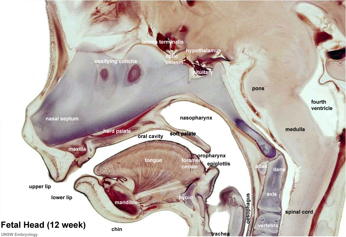 | 1,200 × 821 (186 KB) | Z8600021 (talk | contribs) | |
| 12:23, 18 March 2012 |  | 1,200 × 821 (185 KB) | Z8600021 (talk | contribs) | ||
| 12:17, 18 March 2012 |  | 965 × 660 (120 KB) | Z8600021 (talk | contribs) | ||
| 11:11, 18 March 2012 | 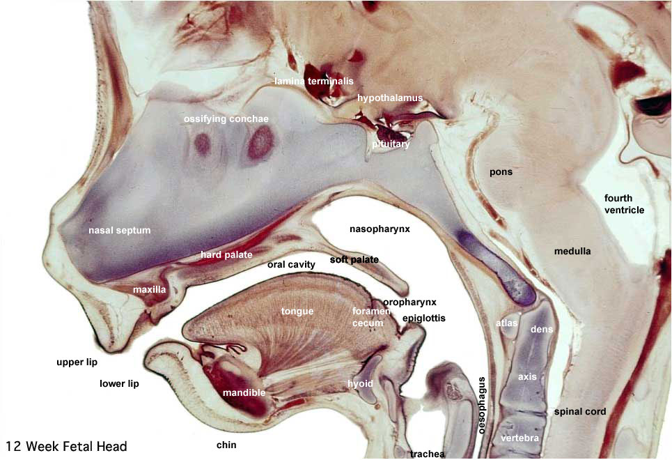 | 965 × 660 (118 KB) | Z8600021 (talk | contribs) | ==Human Fetal Head Week 12== Selected medial head (12 weeks) view showing key features of head musculoskeletal and neurological development. Note extensive nasal cartilage, nasal conchae, pituitary, secondary palate, oral cavity, tongue, mandible, hyoid, |
You cannot overwrite this file.
File usage
The following 14 pages use this file:
- AACP Meeting 2013 - Face Embryology
- ANAT2341 Lab 11 - Embryo to Fetus
- ANAT2341 Lab 6 - Fetal
- BGDA Practical 12 - Second Trimester
- BGDB Face and Ear - Fetal
- BGDB Gastrointestinal - Fetal
- Draft 2016
- Fetal Development - 12 Weeks
- Gastrointestinal Tract - Mouth Development
- Gastrointestinal Tract - Oesophagus Development
- Lecture - Respiratory Development
- Musculoskeletal System - Skull Development
- Neural - Pons Development
- SH Lecture - Respiratory System Development