File:Fetal head section 01.jpg: Difference between revisions
(==Human Fetal Head Week 12== Selected medial head (12 weeks) view showing key features of head musculoskeletal and neurological development. Note extensive nasal cartilage, nasal conchae, pituitary, secondary palate, oral cavity, tongue, mandible, hyoid,) |
|||
| Line 2: | Line 2: | ||
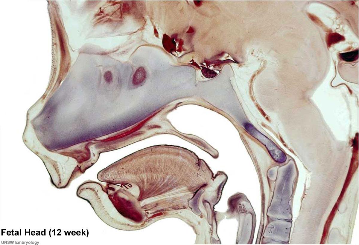
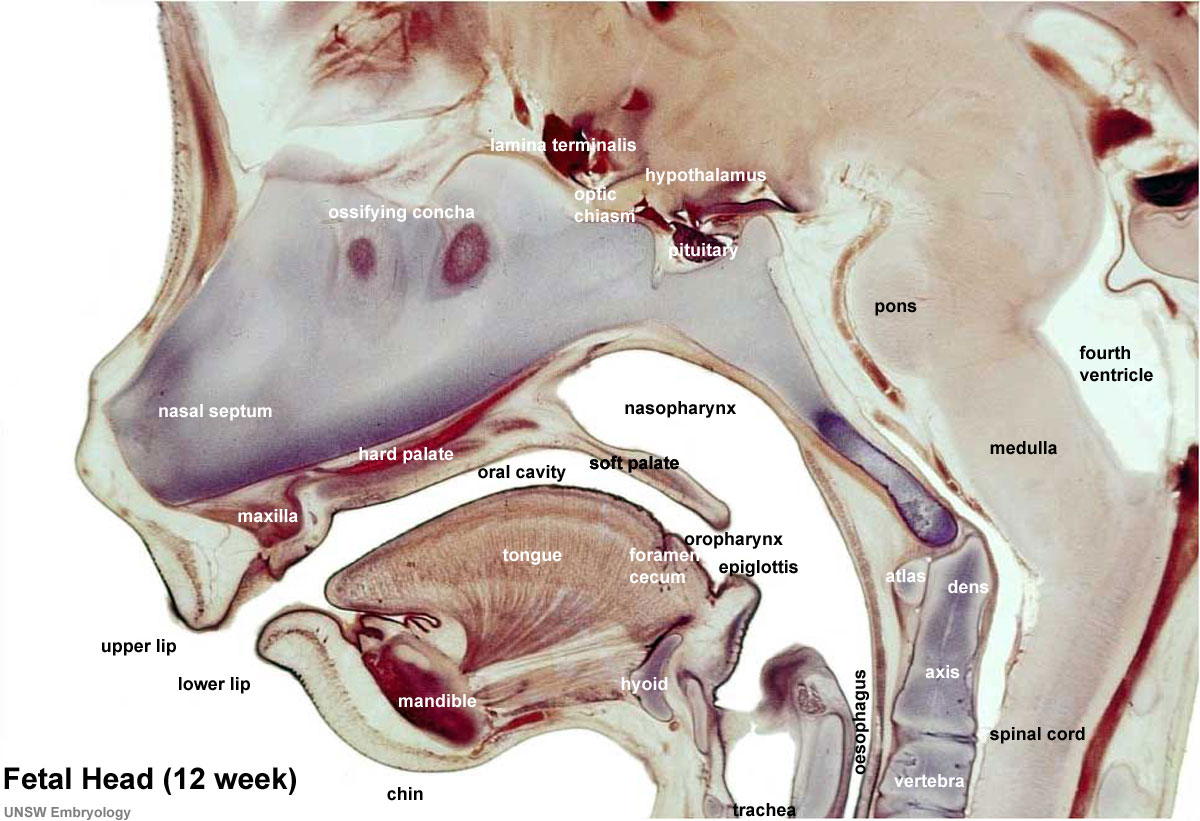
Selected medial head (12 weeks) view showing key features of head musculoskeletal and neurological development. Note extensive nasal cartilage, nasal conchae, pituitary, secondary palate, oral cavity, tongue, mandible, hyoid, choana, oropharynx. Also note the developing tongue musculature and its mandibular attachment site. | Selected medial head (12 weeks) view showing key features of head musculoskeletal and neurological development. Note extensive nasal cartilage, nasal conchae, pituitary, secondary palate, oral cavity, tongue, mandible, hyoid, choana, oropharynx. Also note the developing tongue musculature and its mandibular attachment site. | ||
:'''Image Links:''' [[File:Fetal head section.jpg|unlabeled version]] | [[File:Fetal head section 01.jpg|labeled version]] | [[Musculoskeletal_System_-_Skull_Development|Skull_Development]] | [[Head Development]] | |||
This mid-line section through the fetal head shows features of the developing skull and the brain, face and mouth. | This mid-line section through the fetal head shows features of the developing skull and the brain, face and mouth. | ||
Revision as of 12:08, 18 March 2012
Human Fetal Head Week 12
Selected medial head (12 weeks) view showing key features of head musculoskeletal and neurological development. Note extensive nasal cartilage, nasal conchae, pituitary, secondary palate, oral cavity, tongue, mandible, hyoid, choana, oropharynx. Also note the developing tongue musculature and its mandibular attachment site.
- Image Links:
 |
|  | Skull_Development | Head Development


This mid-line section through the fetal head shows features of the developing skull and the brain, face and mouth.
Neural
- developing brain and brainstem.
- lamina terminalis (site of anterior neuropore closure).
- fourth ventricle.
- developing pituitary sitting in the sella turcica.
Musculoskeletal
- cartilage - septum of the nose.
- bone - ossifying nasal concha.
- bone - palate roof of mouth.
- cartilage - soft palate back of mouth.
- cartilage - base of skull and vertebra.
- muscle - tongue, attached to mandible, note foramen cecum.
- bone - mandible.
- Links: Image - Head histology | Image - Head medial cartilage/bone | Image - Head lateral cartilage/bone | Head Development | Gastrointestinal Tract Development | Skull Development | Bone Development
Source: Prof Virginia Diewert Original File Name: 12weekhead.jpg
Cite this page: Hill, M.A. (2024, April 16) Embryology Fetal head section 01.jpg. Retrieved from https://embryology.med.unsw.edu.au/embryology/index.php/File:Fetal_head_section_01.jpg
- © Dr Mark Hill 2024, UNSW Embryology ISBN: 978 0 7334 2609 4 - UNSW CRICOS Provider Code No. 00098G
File history
Click on a date/time to view the file as it appeared at that time.
| Date/Time | Thumbnail | Dimensions | User | Comment | |
|---|---|---|---|---|---|
| current | 13:07, 18 March 2012 | | 1,200 × 821 (186 KB) | Z8600021 (talk | contribs) | |
| 12:23, 18 March 2012 |  | 1,200 × 821 (185 KB) | Z8600021 (talk | contribs) | ||
| 12:17, 18 March 2012 |  | 965 × 660 (120 KB) | Z8600021 (talk | contribs) | ||
| 11:11, 18 March 2012 | 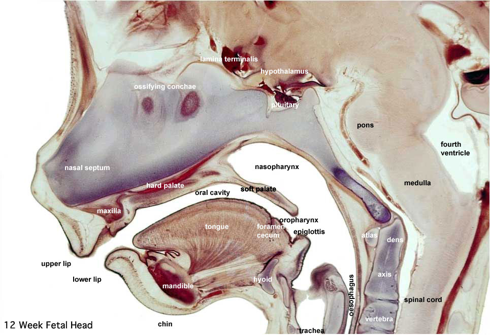 | 965 × 660 (118 KB) | Z8600021 (talk | contribs) | ==Human Fetal Head Week 12== Selected medial head (12 weeks) view showing key features of head musculoskeletal and neurological development. Note extensive nasal cartilage, nasal conchae, pituitary, secondary palate, oral cavity, tongue, mandible, hyoid, |
You cannot overwrite this file.
File usage
The following 14 pages use this file:
- AACP Meeting 2013 - Face Embryology
- ANAT2341 Lab 11 - Embryo to Fetus
- ANAT2341 Lab 6 - Fetal
- BGDA Practical 12 - Second Trimester
- BGDB Face and Ear - Fetal
- BGDB Gastrointestinal - Fetal
- Draft 2016
- Fetal Development - 12 Weeks
- Gastrointestinal Tract - Mouth Development
- Gastrointestinal Tract - Oesophagus Development
- Lecture - Respiratory Development
- Musculoskeletal System - Skull Development
- Neural - Pons Development
- SH Lecture - Respiratory System Development