File:Fetal head section 01.jpg: Difference between revisions
From Embryology
| Line 4: | Line 4: | ||
{| | {| | ||
| valign=top|200px| | | valign=top|width=200px| | ||
===Neural System=== | ===Neural System=== | ||
* [[Neural_System_Development|central nervous system]]. | * [[Neural_System_Development|central nervous system]]. | ||
Revision as of 13:22, 16 January 2016
Human Fetal Head Week 12
This mid-line section through the fetal head (12 weeks) shows features of the developing skull and the brain, face and mouth.
width=200px|
Neural System
|
Endocrine
|
Skeletal System
|
Muscle |
- 12 Week Images: Sagittal unlabeled | Sagittal labeled | Sagittal medial view | Sagittal lateral view | Pituitary unlabeled | Pituitary labeled | Tongue | Skull Development | Head Development
Image Source: Prof Virginia Diewert
Cite this page: Hill, M.A. (2024, April 16) Embryology Fetal head section 01.jpg. Retrieved from https://embryology.med.unsw.edu.au/embryology/index.php/File:Fetal_head_section_01.jpg
- © Dr Mark Hill 2024, UNSW Embryology ISBN: 978 0 7334 2609 4 - UNSW CRICOS Provider Code No. 00098G
File history
Click on a date/time to view the file as it appeared at that time.
| Date/Time | Thumbnail | Dimensions | User | Comment | |
|---|---|---|---|---|---|
| current | 13:07, 18 March 2012 | 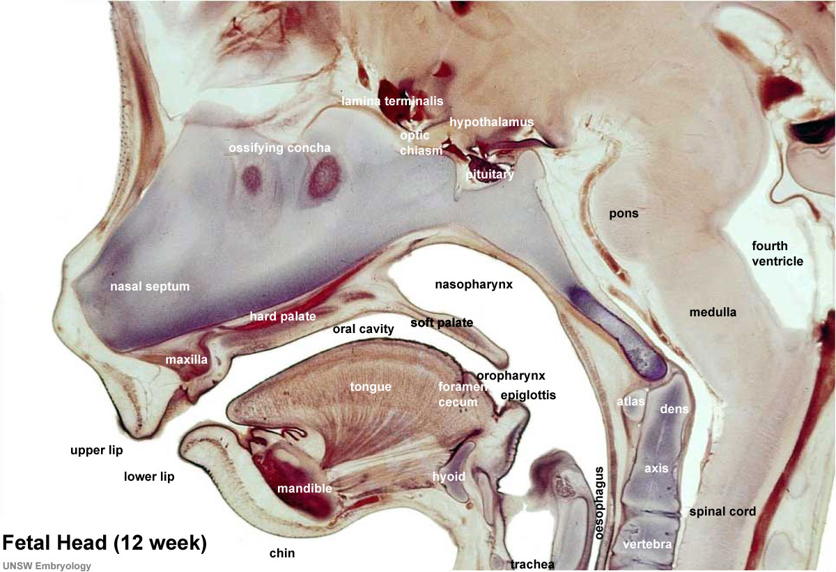 | 1,200 × 821 (186 KB) | Z8600021 (talk | contribs) | |
| 12:23, 18 March 2012 |  | 1,200 × 821 (185 KB) | Z8600021 (talk | contribs) | ||
| 12:17, 18 March 2012 |  | 965 × 660 (120 KB) | Z8600021 (talk | contribs) | ||
| 11:11, 18 March 2012 | 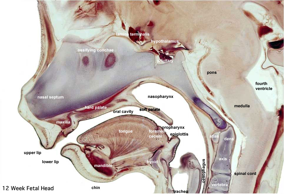 | 965 × 660 (118 KB) | Z8600021 (talk | contribs) | ==Human Fetal Head Week 12== Selected medial head (12 weeks) view showing key features of head musculoskeletal and neurological development. Note extensive nasal cartilage, nasal conchae, pituitary, secondary palate, oral cavity, tongue, mandible, hyoid, |
You cannot overwrite this file.
File usage
The following 14 pages use this file:
- AACP Meeting 2013 - Face Embryology
- ANAT2341 Lab 11 - Embryo to Fetus
- ANAT2341 Lab 6 - Fetal
- BGDA Practical 12 - Second Trimester
- BGDB Face and Ear - Fetal
- BGDB Gastrointestinal - Fetal
- Draft 2016
- Fetal Development - 12 Weeks
- Gastrointestinal Tract - Mouth Development
- Gastrointestinal Tract - Oesophagus Development
- Lecture - Respiratory Development
- Musculoskeletal System - Skull Development
- Neural - Pons Development
- SH Lecture - Respiratory System Development