File:Fetal head section.jpg
From Embryology

Size of this preview: 800 × 547 pixels. Other resolution: 1,200 × 821 pixels.
Original file (1,200 × 821 pixels, file size: 167 KB, MIME type: image/jpeg)
Selected medial head view showing key features of head musculoskeletal and neurological development. Note extensive nasal cartilage, nasal conchae, pituitary, secondary palate, oral cavity, tongue, mandible, hyoid, choana, oropharynx.
Also note the developing tongue musculature and its mandibular attachment site.
Original File Name: 12weekhead.jpg
http://embryology.med.unsw.edu.au/wwwhuman/Hum12wk/Hum12wk.htm
File history
Click on a date/time to view the file as it appeared at that time.
| Date/Time | Thumbnail | Dimensions | User | Comment | |
|---|---|---|---|---|---|
| current | 12:24, 18 March 2012 | | 1,200 × 821 (167 KB) | Z8600021 (talk | contribs) | |
| 12:18, 18 March 2012 | 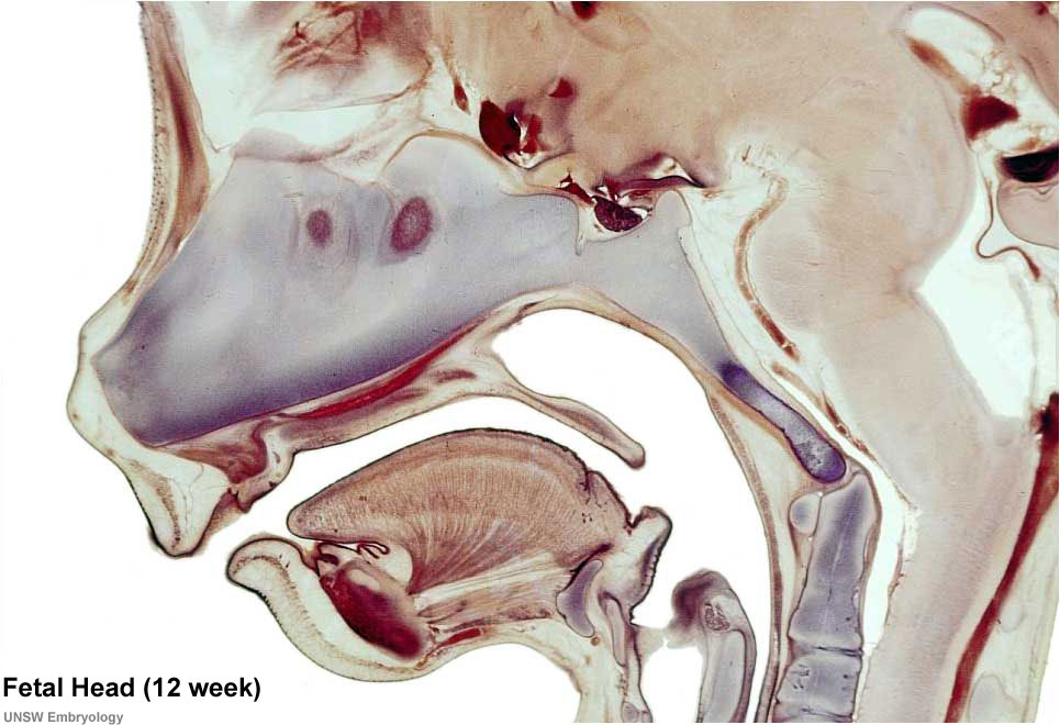 | 965 × 660 (107 KB) | Z8600021 (talk | contribs) | ||
| 15:41, 13 September 2009 |  | 965 × 660 (63 KB) | S8600021 (talk | contribs) | Selected medial head view showing key features of head musculoskeletal and neurological development. Note extensive nasal cartilage, nasal conchae, pituitary, secondary palate, oral cavity, tongue, mandible, hyoid, choana, oropharynx. Also note the devel |
You cannot overwrite this file.
File usage
The following 26 pages use this file:
- 2009 Lecture 13
- 2010 Foundations Lecture - Introduction to Human Development
- 2010 Lab 6
- 2010 Lecture 13
- 2011 Lab 6 - Fetal
- AACP Meeting 2013 - Face Embryology
- ANAT2241 Bone, Bone Formation and Joints
- ANAT2341 Lab 6 - Fetal
- Abnormal Development - Fetal Growth Restriction
- BGDB Face and Ear - Fetal
- Bone Development
- Bone Histology
- Cartilage Histology
- Fetal Development
- Fetal Development - 12 Weeks
- Foundations Lecture - Introduction to Human Development
- Head Development
- Joint Development - Temporomandibular Joint
- Lecture - Musculoskeletal Development
- Musculoskeletal System - Abnormalities
- Musculoskeletal System - Axial Skeleton Development
- Musculoskeletal System - Bone Development
- Musculoskeletal System - Cartilage Development
- Musculoskeletal System - Joint Development
- Musculoskeletal System Development
- Pre-Medicine Program - Embryology