File:Fetal gonad retinoid receptor expression 01.jpg: Difference between revisions
mNo edit summary |
mNo edit summary |
||
| Line 1: | Line 1: | ||
==Human Fetal Gonad Retinoid Receptor Expression== | ==Human Fetal Gonad Retinoid Receptor Expression== | ||
{{ | Second trimester human {{fetal}} {{testis}} and {{ovary}}. | ||
{| | {| | ||
Latest revision as of 09:38, 15 July 2018
Human Fetal Gonad Retinoid Receptor Expression
Second trimester human fetal testis and ovary.
|
|
|
|
The widespread nuclear localization of RA receptors in testis suggests cells of all types (including germ cells) are exposed to RA signals. |
|
Magnification: 400× (A, B), 1000× (C–F).
Reference
Childs AJ, Cowan G, Kinnell HL, Anderson RA & Saunders PT. (2011). Retinoic Acid signalling and the control of meiotic entry in the human fetal gonad. PLoS ONE , 6, e20249. PMID: 21674038 DOI.
Copyright
© 2011 Childs et al. This is an open-access article distributed under the terms of the Creative Commons Attribution License, which permits unrestricted use, distribution, and reproduction in any medium, provided the original author and source are credited.
Text edited from original figure legend. Figure 3. doi:info:doi/10.1371/journal.pone.0020249.g003
Cite this page: Hill, M.A. (2024, April 18) Embryology Fetal gonad retinoid receptor expression 01.jpg. Retrieved from https://embryology.med.unsw.edu.au/embryology/index.php/File:Fetal_gonad_retinoid_receptor_expression_01.jpg
- © Dr Mark Hill 2024, UNSW Embryology ISBN: 978 0 7334 2609 4 - UNSW CRICOS Provider Code No. 00098G
File history
Click on a date/time to view the file as it appeared at that time.
| Date/Time | Thumbnail | Dimensions | User | Comment | |
|---|---|---|---|---|---|
| current | 17:48, 21 May 2012 | 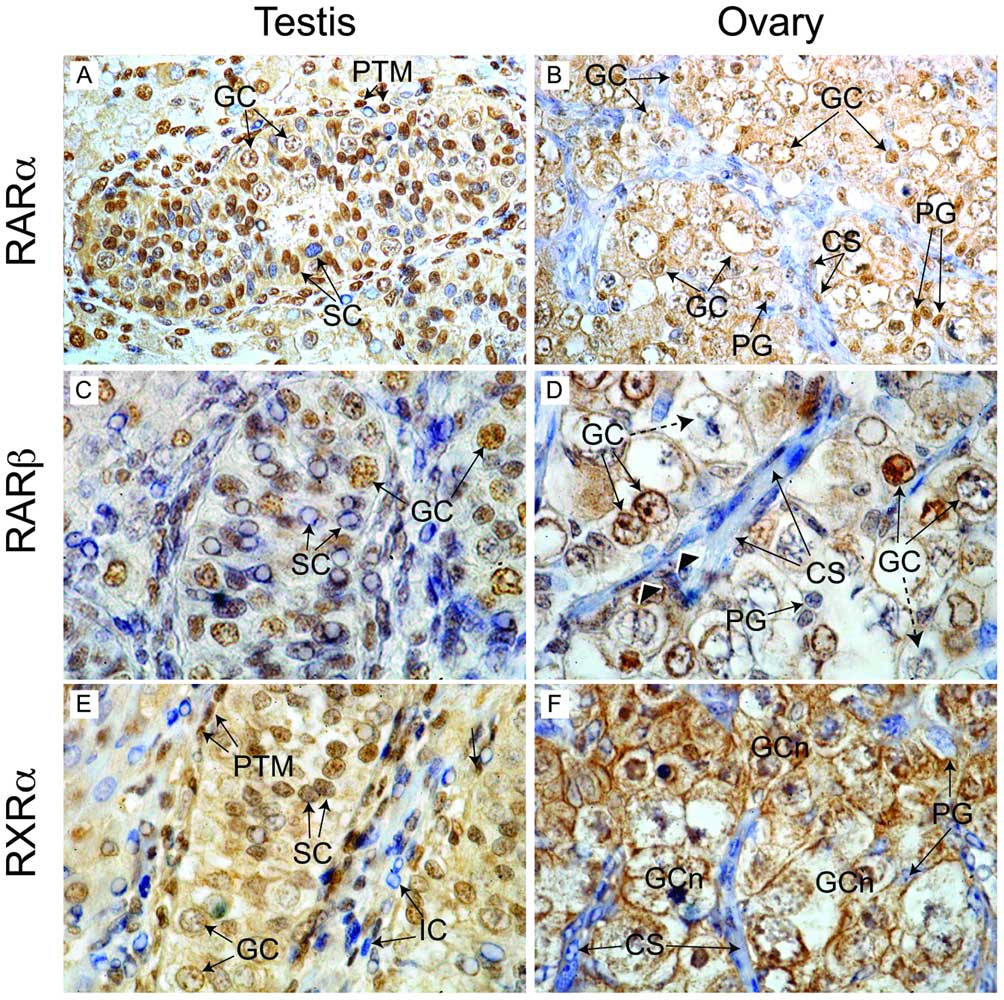 | 1,004 × 1,000 (226 KB) | Z8600021 (talk | contribs) | Immunohistochemical localisation of retinoid receptor expression in the human fetal gonad. In the second trimester human fetal testis (A) RARα staining was detected in germ cell (GC) and peritubular myoid (PTM) nuclei. Two populations of Sertoli cells ( |
You cannot overwrite this file.
File usage
The following 2 pages use this file: